 Movie
Movie Controller


+ Open data

- Basic information
| Entry |  | |||||||||
|---|---|---|---|---|---|---|---|---|---|---|
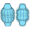
| Title | Cryo-EM structure of RNF170-Erlin1-Erlin2 | |||||||||
 Map data Map data | ||||||||||
 Sample Sample |
| |||||||||
 Keywords Keywords | Complex / STRUCTURAL PROTEIN | |||||||||
| Function / homology |  Function and homology information Function and homology informationregulation of cholesterol biosynthetic process / Signaling by plasma membrane FGFR1 fusions / SREBP signaling pathway / negative regulation of cholesterol biosynthetic process / negative regulation of fatty acid biosynthetic process / cholesterol binding / cholesterol metabolic process / ERAD pathway / Signaling by FGFR1 in disease / Defective CFTR causes cystic fibrosis ...regulation of cholesterol biosynthetic process / Signaling by plasma membrane FGFR1 fusions / SREBP signaling pathway / negative regulation of cholesterol biosynthetic process / negative regulation of fatty acid biosynthetic process / cholesterol binding / cholesterol metabolic process / ERAD pathway / Signaling by FGFR1 in disease / Defective CFTR causes cystic fibrosis / AMPK-induced ERAD and lysosome mediated degradation of PD-L1(CD274) / ABC-family protein mediated transport / membrane raft / ubiquitin protein ligase binding / endoplasmic reticulum membrane / endoplasmic reticulum / protein-containing complex / plasma membrane Similarity search - Function | |||||||||
| Biological species |  Homo sapiens (human) Homo sapiens (human) | |||||||||
| Method | single particle reconstruction / cryo EM / Resolution: 3.05 Å | |||||||||
 Authors Authors | Qian HW / Jia XX | |||||||||
| Funding support |  China, 1 items China, 1 items
| |||||||||
 Citation Citation | Journal: J Mol Cell Biol / Year: 2026 Title: Structural insights into the organization of the human Erlin complex. Authors: Xiaoxiao Jia / Guoyun Liu / Haiwen Li / Hongwu Qian / Abstract: The endoplasmic reticulum lipid raft proteins (Erlins) belong to the stomatin-prohibitin-flotillin-HflC/K (SPFH) family and form highly oligomeric platforms that mediate the degradation of activated ...The endoplasmic reticulum lipid raft proteins (Erlins) belong to the stomatin-prohibitin-flotillin-HflC/K (SPFH) family and form highly oligomeric platforms that mediate the degradation of activated inositol 1,4,5-trisphosphate receptors by facilitating their interaction with the E3 ligase RNF170. However, the molecular mechanisms underlying this process remain unclear. Here, we successfully reconstituted the Erlin1-Erlin2 complex and its complex with RNF170 by overexpressing these components in HEK293F cells. We also isolated the Erlin2 oligomer by solely expressing Erlin2 in the cells. Using cryo-electron microscopy, we determined the structures of the Erlin1-Erlin2 complex, the Erlin1-Erlin2-RNF170 complex, and the Erlin2 oligomer at resolutions of 3.29 Å, 3.05 Å, and 2.12 Å, respectively. Both the Erlin1-Erlin2 complex and the Erlin2 oligomer exhibit similar cage-like architectures, with the Erlin1-Erlin2 complex containing 13 pairs of Erlin1 and Erlin2 subunits, whereas the Erlin2 oligomer comprises 26 Erlin2. Although RNF170 was clearly identified during protein purification, it was invisible in the final 3D reconstruction, suggesting a high degree of flexibility between RNF170 and the Erlin complex. Multiple water molecules were identified in the Erlin2 oligomer, underscoring their critical roles in facilitating the high degree of oligomerization of the Erlin2 complex. Taken together, our structural investigation elucidates the molecular basis for the assembly of the Erlin complex and provides a framework for further investigation. | |||||||||
| History |
|
- Structure visualization
| Supplemental images |
|---|

- Downloads & links
-EMDB archive
| Map data | emd_65914.map.gz | 167.9 MB | EMDB map data format | |
|---|---|---|---|---|
| Header (meta data) | emd-65914-v30.xmlemd-65914.xml | 17.2 KB 17.2 KB | Display Display | EMDB header |
| Images |  emd_65914.png emd_65914.png | 99.3 KB | ||
| Filedesc metadata | emd-65914.cif.gz | 5.8 KB | ||
| Others | emd_65914_half_map_1.map.gzemd_65914_half_map_2.map.gz | 164.6 MB 164.6 MB | ||
| Archive directory |  https://data.pdbj.org/pub/emdb/structures/EMD-65914ftp://data.pdbj.org/pub/emdb/structures/EMD-65914 https://data.pdbj.org/pub/emdb/structures/EMD-65914ftp://data.pdbj.org/pub/emdb/structures/EMD-65914 | HTTPS FTP |
-Related structure data
| Related structure data |  9we7MC  9wffC M: atomic model generated by this map C: citing same article ( |
|---|---|
| Similar structure data |


-Links
| EMDB pages | EMDB (EBI/PDBe) / EMDataResource |
|---|---|
| Related items in Molecule of the Month |


-Map
| File | Download / File: emd_65914.map.gz / Format: CCP4 / Size: 178 MB / Type: IMAGE STORED AS FLOATING POINT NUMBER (4 BYTES) | ||||||||||||||||||||||||||||||||||||
|---|---|---|---|---|---|---|---|---|---|---|---|---|---|---|---|---|---|---|---|---|---|---|---|---|---|---|---|---|---|---|---|---|---|---|---|---|---|
| Projections & slices | Image control
Images are generated by Spider. | ||||||||||||||||||||||||||||||||||||
| Voxel size | X=Y=Z: 1.07 Å | ||||||||||||||||||||||||||||||||||||
| Density |
| ||||||||||||||||||||||||||||||||||||
| Symmetry | Space group: 1 | ||||||||||||||||||||||||||||||||||||
| Details | EMDB XML:
|
 Z (Sec.)
 Y (Row.)
 X (Col.)


-Supplemental data
-Half map: #2
| File | emd_65914_half_map_1.map | ||||||||||||
|---|---|---|---|---|---|---|---|---|---|---|---|---|---|
| Projections & Slices |
| ||||||||||||
| Density Histograms |


-Half map: #1
| File | emd_65914_half_map_2.map | ||||||||||||
|---|---|---|---|---|---|---|---|---|---|---|---|---|---|
| Projections & Slices |
| ||||||||||||
| Density Histograms |


- Sample components
-Entire : Cryo-EM structure of A
| Entire | Name: Cryo-EM structure of A |
|---|---|
| Components |
|
-Supramolecule #1: Cryo-EM structure of A
| Supramolecule | Name: Cryo-EM structure of A / type: complex / ID: 1 / Parent: 0 / Macromolecule list: #1-#2 |
|---|---|
| Source (natural) | Organism: Homo sapiens (human) |
-Macromolecule #1: Erlin-1
| Macromolecule | Name: Erlin-1 / type: protein_or_peptide / ID: 1 / Number of copies: 13 / Enantiomer: LEVO |
|---|---|
| Source (natural) | Organism: Homo sapiens (human) |
| Molecular weight | Theoretical: 31.840676 KDa |
| Recombinant expression | Organism: Homo sapiens (human) |
| Sequence | String: YASIHKIEEG HLAVYYRGGA LLTSPSGPGY HIMLPFITTF RSVQTTLQTD EVKNVPCGTS GGVMIYIDRI EVVNMLAPYA VFDIVRNYT ADYDKTLIFN KIHHELNQFC SAHTLQEVYI ELFDQIDENL KQALQKDLNL MAPGLTIQAV RVTKPKIPEA I RRNFELME ...String: YASIHKIEEG HLAVYYRGGA LLTSPSGPGY HIMLPFITTF RSVQTTLQTD EVKNVPCGTS GGVMIYIDRI EVVNMLAPYA VFDIVRNYT ADYDKTLIFN KIHHELNQFC SAHTLQEVYI ELFDQIDENL KQALQKDLNL MAPGLTIQAV RVTKPKIPEA I RRNFELME AEKTKLLIAA QKQKVVEKEA ETERKKAVIE AEKIAQVAKI RFQQKVMEKE TEKRISEIED AAFLAREKAK AD AEYYAAH KYATSNKHKL TPEYLELKKY QAIASNSKIY F UniProtKB: Erlin-1 |
-Macromolecule #2: Erlin-2
| Macromolecule | Name: Erlin-2 / type: protein_or_peptide / ID: 2 / Number of copies: 13 / Enantiomer: LEVO |
|---|---|
| Source (natural) | Organism: Homo sapiens (human) |
| Molecular weight | Theoretical: 31.349125 KDa |
| Recombinant expression | Organism: Homo sapiens (human) |
| Sequence | String: SAVHKIEEGH IGVYYRGGAL LTSTSGPGFH LMLPFITSYK SVQTTLQTDE VKNVPCGTSG GVMIYFDRIE VVNFLVPNAV YDIVKNYTA DYDKALIFNK IHHELNQFCS VHTLQEVYIE LFDQIDENLK LALQQDLTSM APGLVIQAVR VTKPNIPEAI R RNYELMES ...String: SAVHKIEEGH IGVYYRGGAL LTSTSGPGFH LMLPFITSYK SVQTTLQTDE VKNVPCGTSG GVMIYFDRIE VVNFLVPNAV YDIVKNYTA DYDKALIFNK IHHELNQFCS VHTLQEVYIE LFDQIDENLK LALQQDLTSM APGLVIQAVR VTKPNIPEAI R RNYELMES EKTKLLIAAQ KQKVVEKEAE TERKKALIEA EKVAQVAEIT YGQKVMEKET EKKISEIEDA AFLAREKAKA DA ECYTAMK IAEANKLKLT PEYLQLMKYK AIASNSKIYF UniProtKB: Erlin-2 |
-Macromolecule #3: 2-acetamido-2-deoxy-beta-D-glucopyranose
| Macromolecule | Name: 2-acetamido-2-deoxy-beta-D-glucopyranose / type: ligand / ID: 3 / Number of copies: 26 / Formula: NAG |
|---|---|
| Molecular weight | Theoretical: 221.208 Da |
| Chemical component information |  ChemComp-NAG: |
-Experimental details
-Structure determination
| Method | cryo EM |
|---|---|
 Processing Processing | single particle reconstruction |
| Aggregation state | particle |
-Sample preparation
| Buffer | pH: 8 |
|---|---|
| Vitrification | Cryogen name: ETHANE |
- Electron microscopy
| Microscope | FEI POLARA 300 |
|---|---|
| Image recording | Film or detector model: GATAN K3 (6k x 4k) / Average electron dose: 50.0 e/Å2 |
| Electron beam | Acceleration voltage: 300 kV / Electron source:  FIELD EMISSION GUN FIELD EMISSION GUN |
| Electron optics | Illumination mode: FLOOD BEAM / Imaging mode: BRIGHT FIELD / Nominal defocus max: 1.7 µm / Nominal defocus min: 1.5 µm |
| Experimental equipment | 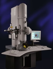 Model: Tecnai Polara / Image courtesy: FEI Company |
