 Movie
Movie Controller


+ Open data

- Basic information
| Entry |  | |||||||||
|---|---|---|---|---|---|---|---|---|---|---|
| Title | Tspan-7 Tetramer Structure in Retraction Fiber | |||||||||
 Map data Map data | Author claim: Regarding the FSC curve, we carefully re-examined the refinement and the two independent half maps. We did not find evidence of half-set contamination during refinement. In our case, the reconstruction was performed using helical local refinement of the minimal repeat unit. For this type of reconstruction, the calculated FSC may remain slightly above zero at high spatial frequencies due to the combination of helical symmetry, local refinement, masking, and interpolation procedures, rather than indicating correlation introduced by mixing the two half datasets. The FSC crosses the 0.143 criterion at 6.63 A as expected, and the residual correlation at higher frequencies remains very low (approximately 0.02-0.03) without any abnormal increase, which we believe is consistent with residual background correlation rather than half-map contamination. We have carefully verified that the refinement followed gold-standard procedures using independent half datasets throughout the reconstruction. | |||||||||
 Sample Sample |
| |||||||||
 Keywords Keywords | Membrane curvature / cell protrusion / helical structure / MEMBRANE PROTEIN | |||||||||
| Function / homology |  Function and homology information Function and homology informationTrafficking of GluR2-containing AMPA receptors / Cell surface interactions at the vascular wall / plasma membrane Similarity search - Function | |||||||||
| Biological species |   Rattus norvegicus (Norway rat) Rattus norvegicus (Norway rat) | |||||||||
| Method | single particle reconstruction / cryo EM / Resolution: 6.63 Å | |||||||||
 Authors Authors | Jia X / Wang DJ / Li XP / Liu N / Yu L / Wang HW | |||||||||
| Funding support |  China, 1 items China, 1 items
| |||||||||
 Citation Citation | Journal: Vita / Year: 2026 Title: Polymerization of tetraspanin 7 into helical transmembrane skeletons for Authors: Wang D / Jia X / Dharan R / Ren J / Zheng Y / Li X / Huang M / Xu K / Zhang Q / Sho T / Liu S / Yang F / Zhang QC / Sorkin R / Liu N / Wang HW / Yu L | |||||||||
| History |
|
- Structure visualization
| Supplemental images |
|---|

- Downloads & links
-EMDB archive
| Map data | emd_65484.map.gz | 59 MB | EMDB map data format | |
|---|---|---|---|---|
| Header (meta data) | emd-65484-v30.xmlemd-65484.xml | 15.1 KB 15.1 KB | Display Display | EMDB header |
| Images |  emd_65484.png emd_65484.png | 51.7 KB | ||
| Masks | emd_65484_msk_1.map | 64 MB | Mask map | |
| Filedesc metadata | emd-65484.cif.gz | 4.5 KB | ||
| Others | emd_65484_half_map_1.map.gzemd_65484_half_map_2.map.gz | 56.9 MB 56.9 MB | ||
| Archive directory |  http://ftp.pdbj.org/pub/emdb/structures/EMD-65484ftp://ftp.pdbj.org/pub/emdb/structures/EMD-65484 http://ftp.pdbj.org/pub/emdb/structures/EMD-65484ftp://ftp.pdbj.org/pub/emdb/structures/EMD-65484 | HTTPS FTP |
-Related structure data
| Related structure data |  9w2dMC  9w2bC M: atomic model generated by this map C: citing same article ( |
|---|---|
| Similar structure data |


-Links
| EMDB pages | EMDB (EBI/PDBe) / EMDataResource |
|---|---|
| Related items in Molecule of the Month |
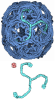
-Map
| File | Download / File: emd_65484.map.gz / Format: CCP4 / Size: 64 MB / Type: IMAGE STORED AS FLOATING POINT NUMBER (4 BYTES) | ||||||||||||||||||||||||||||||||||||
|---|---|---|---|---|---|---|---|---|---|---|---|---|---|---|---|---|---|---|---|---|---|---|---|---|---|---|---|---|---|---|---|---|---|---|---|---|---|
| Annotation | Author claim: Regarding the FSC curve, we carefully re-examined the refinement and the two independent half maps. We did not find evidence of half-set contamination during refinement. In our case, the reconstruction was performed using helical local refinement of the minimal repeat unit. For this type of reconstruction, the calculated FSC may remain slightly above zero at high spatial frequencies due to the combination of helical symmetry, local refinement, masking, and interpolation procedures, rather than indicating correlation introduced by mixing the two half datasets. The FSC crosses the 0.143 criterion at 6.63 A as expected, and the residual correlation at higher frequencies remains very low (approximately 0.02-0.03) without any abnormal increase, which we believe is consistent with residual background correlation rather than half-map contamination. We have carefully verified that the refinement followed gold-standard procedures using independent half datasets throughout the reconstruction. | ||||||||||||||||||||||||||||||||||||
| Projections & slices | Image control
Images are generated by Spider. | ||||||||||||||||||||||||||||||||||||
| Voxel size | X=Y=Z: 1.36 Å | ||||||||||||||||||||||||||||||||||||
| Density |
| ||||||||||||||||||||||||||||||||||||
| Symmetry | Space group: 1 | ||||||||||||||||||||||||||||||||||||
| Details | EMDB XML:
|
 Z (Sec.)
 Y (Row.)
 X (Col.)


-Supplemental data
-Mask #1
| File | emd_65484_msk_1.map | ||||||||||||
|---|---|---|---|---|---|---|---|---|---|---|---|---|---|
| Projections & Slices |
| ||||||||||||
| Density Histograms |


-Half map: #1
| File | emd_65484_half_map_1.map | ||||||||||||
|---|---|---|---|---|---|---|---|---|---|---|---|---|---|
| Projections & Slices |
| ||||||||||||
| Density Histograms |


-Half map: #2
| File | emd_65484_half_map_2.map | ||||||||||||
|---|---|---|---|---|---|---|---|---|---|---|---|---|---|
| Projections & Slices |
| ||||||||||||
| Density Histograms |


- Sample components
-Entire : Tspan-7 Tetramer in membrane tube
| Entire | Name: Tspan-7 Tetramer in membrane tube |
|---|---|
| Components |
|
-Supramolecule #1: Tspan-7 Tetramer in membrane tube
| Supramolecule | Name: Tspan-7 Tetramer in membrane tube / type: organelle_or_cellular_component / ID: 1 / Parent: 0 |
|---|---|
| Source (natural) | Organism: Rattus norvegicus (Norway rat) |
-Experimental details
-Structure determination
| Method | cryo EM |
|---|---|
 Processing Processing | single particle reconstruction |
| Aggregation state | particle |
-Sample preparation
| Buffer | pH: 7.4 |
|---|---|
| Vitrification | Cryogen name: ETHANE / Chamber humidity: 100 % / Chamber temperature: 281.15 K |
- Electron microscopy
| Microscope | TFS KRIOS |
|---|---|
| Image recording | Film or detector model: GATAN K3 BIOQUANTUM (6k x 4k) / Average electron dose: 50.0 e/Å2 |
| Electron beam | Acceleration voltage: 300 kV / Electron source:  FIELD EMISSION GUN FIELD EMISSION GUN |
| Electron optics | Illumination mode: FLOOD BEAM / Imaging mode: BRIGHT FIELD / Nominal defocus max: 1.8 µm / Nominal defocus min: 1.2 µm |
| Experimental equipment |  Model: Titan Krios / Image courtesy: FEI Company |
