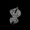
[English] 日本語
Yorodumi
- EMDB-65259: Cryo-EM structure of the dimeric Elapor1 mutant -

+
Open data


ID or keywords:

Loading...

-
Basic information

Entry
Database: EMDB / ID: EMD-65259
TitleCryo-EM structure of the dimeric Elapor1 mutant
Map data
Sample
  • Complex: Elapor1 mutant
    • Protein or peptide: Endosome/lysosome-associated apoptosis and autophagy regulator 1
  • Ligand: 2-acetamido-2-deoxy-beta-D-glucopyranose
KeywordsTethering factor / MEMBRANE PROTEIN
Function / homology
Function and homology information


positive regulation of autophagosome assembly / autophagosome assembly / cellular response to starvation / trans-Golgi network / late endosome / lysosome / plasma membrane
Similarity search - Function
EIG121-like / : / : / : / : / : / ELAPOR1-like, galactose binding domain / ELAPOR1-like, galactose-binding domain / ELAPOR1-like, mannose 6-phosphate receptor homology domain / ELAPOR1-like, C-terminal domain ...EIG121-like / : / : / : / : / : / ELAPOR1-like, galactose binding domain / ELAPOR1-like, galactose-binding domain / ELAPOR1-like, mannose 6-phosphate receptor homology domain / ELAPOR1-like, C-terminal domain / ELAPOR1-like, TNF receptor-like / Mannose-6-phosphate receptor binding domain superfamily / MRH domain / MRH domain profile. / Putative ephrin-receptor like / Growth factor receptor cysteine-rich domain superfamily
Similarity search - Domain/homology
Endosome/lysosome-associated apoptosis and autophagy regulator 1
Similarity search - Component
Biological speciesMus musculus (house mouse)
Methodsingle particle reconstruction / cryo EM / Resolution: 3.6 Å
AuthorsMa J / Zheng S
Funding support China, 1 items
OrganizationGrant numberCountry
National Science Foundation (NSF, China) China
CitationJournal: To Be Published
Title: ELAPOR1 is A Copper-Dependent Tethering Factor Driving Proacrosomal Vesicle Fusion in Acrosome Biogenesis
Authors: Shao T / Ma J
History
DepositionJul 5, 2025-
Header (metadata) releaseJul 23, 2025-
Map releaseJul 23, 2025-
UpdateJul 23, 2025-
Current statusJul 23, 2025Processing site: PDBc / Status: Released

-
Structure visualization

Supplemental images

Downloads & links

-
Map

FileDownload / File: emd_65259.map.gz / Format: CCP4 / Size: 103 MB / Type: IMAGE STORED AS FLOATING POINT NUMBER (4 BYTES)
Projections & slices

Image control

Size
Brightness
Contrast
Others
AxesZ (Sec.)Y (Row.)X (Col.)
1.09 Å/pix.
x 300 pix.
= 326.1 Å
1.09 Å/pix.
x 300 pix.
= 326.1 Å
1.09 Å/pix.
x 300 pix.
= 326.1 Å

Surface

Projections

Slices (1/3)

Slices (1/2)

Slices (2/3)

Images are generated by Spider.

Voxel sizeX=Y=Z: 1.087 Å
Density
Contour LevelBy AUTHOR: 0.2
Minimum - Maximum-2.4240885 - 3.317913
Average (Standard dev.)-0.0015328772 (±0.05691853)
SymmetrySpace group: 1
Details

EMDB XML:

Map geometry
Axis orderXYZ
Origin000
Dimensions300300300
Spacing300300300
CellA=B=C: 326.1 Å
α=β=γ: 90.0 °

-
Supplemental data

-
Half map: #2

Fileemd_65259_half_map_1.map
Projections & Slices
AxesZYX

Projections

Slices (1/2)
Density Histograms

-
Half map: #1

Fileemd_65259_half_map_2.map
Projections & Slices
AxesZYX

Projections

Slices (1/2)
Density Histograms

-
Sample components

-
Entire : Elapor1 mutant

EntireName: Elapor1 mutant
Components
  • Complex: Elapor1 mutant
    • Protein or peptide: Endosome/lysosome-associated apoptosis and autophagy regulator 1
  • Ligand: 2-acetamido-2-deoxy-beta-D-glucopyranose

-
Supramolecule #1: Elapor1 mutant

SupramoleculeName: Elapor1 mutant / type: complex / ID: 1 / Parent: 0 / Macromolecule list: #1
Source (natural)Organism: Mus musculus (house mouse)

-
Macromolecule #1: Endosome/lysosome-associated apoptosis and autophagy regulator 1

MacromoleculeName: Endosome/lysosome-associated apoptosis and autophagy regulator 1
type: protein_or_peptide / ID: 1 / Number of copies: 2 / Enantiomer: LEVO
Source (natural)Organism: Mus musculus (house mouse)
Molecular weightTheoretical: 110.429422 KDa
Recombinant expressionOrganism: Homo sapiens (human)
SequenceString: MKTIIALSYI FCLVFADYKD DDDKGSHHHH HHSGMGPELA ACKESEYAFE YTACDSTGSR WRVAVPASPG LCTSLPDPVK GTECSFSCN AGEFLDMKDQ SCKPCAEGRY SLGTGIRFDE WDELPHGFAS LSANLEVDDS ISESTENCTS SKWVPRGDYI A SNTDECTA ...String:
MKTIIALSYI FCLVFADYKD DDDKGSHHHH HHSGMGPELA ACKESEYAFE YTACDSTGSR WRVAVPASPG LCTSLPDPVK GTECSFSCN AGEFLDMKDQ SCKPCAEGRY SLGTGIRFDE WDELPHGFAS LSANLEVDDS ISESTENCTS SKWVPRGDYI A SNTDECTA TLMYAVNLKQ SGTVNFEYYY PDSSIIFEFF VQNDQCQPSA DDSRWMKTTE KGWEFHSVEL NRGNNVLYWR TT AFSVWSK VSKPVLVRNI AITGVAYTSE CFPCKPGTYA AKQGSPFCKL CPANYYSNKG ETSCAPCDAD KYSEKGSSTC KVR PACTDK DYFYTHTACD AQGETQLMYK WAIPKICGED LEGAVKLPAS GVKTRCPPCN PGFFKTDNST CEPCPYGSYS NGSD CTHCP AGTEPAVGFE YKWWNTLPSN METTVLSGIN FEYKGLTGWE VAGDHIYTAV GASDNDFMIL TLVVPGFRPP QSVVA DTEN KEVARITFVF ETICSVNCEL YFMVGMNSRT NTPVETWKGT KGKQSYTYTI EENATVSFTW AFQRTTLHET GRKYTN DVA KIYSINVTNV MGGVASYCRP CALEASDLGS SCTSCPAGHY INRDSGTCHL CPSNTILKAH QPYGAQACVP CGPGTKN NK IHSLCYNDCT FSRNTPSRIF NYNFSALAGT VSLAGVPSFT SKGLKYFHHF TLSLCGNQGK KMAVCTDNVT DLRIPDGE A GFSKSVTAYV CQVVIIPSEV MGYKAGVSSQ PVSLADRLVG VSTDMTLEGI VSPVELFHPE TSGIPDIVFF FRSNDVTQS CSSGRSTTIR LRCNPMKAAP GTLRLPSMCS DGTCDGCNFH FLWESVAACP LCSASDYHTF VSSCVAGIQK TTYMWREPKL CSGGISLPE QRVTICKTID FWLKVGISAG TCTAILLTVL TCYFWKKNQK LEYKYSKLVM NATLKDCDLP AADSCAIMEG E DVEDDFIF TSKKSLFGKI KSFTSKRTPD GFDSVPLKTS SGGPDMDM

UniProtKB: Endosome/lysosome-associated apoptosis and autophagy regulator 1

-
Macromolecule #3: 2-acetamido-2-deoxy-beta-D-glucopyranose

MacromoleculeName: 2-acetamido-2-deoxy-beta-D-glucopyranose / type: ligand / ID: 3 / Number of copies: 6 / Formula: NAG
Molecular weightTheoretical: 221.208 Da
Chemical component information

ChemComp-NAG:
2-acetamido-2-deoxy-beta-D-glucopyranose

-
Experimental details

-
Structure determination

Methodcryo EM
Processingsingle particle reconstruction
Aggregation stateparticle

-
Sample preparation

BufferpH: 7.4
VitrificationCryogen name: ETHANE

-
Electron microscopy

MicroscopeFEI TALOS ARCTICA
Image recordingFilm or detector model: GATAN K3 (6k x 4k) / Average electron dose: 50.0 e/Å2
Electron beamAcceleration voltage: 300 kV / Electron source: FIELD EMISSION GUN
Electron opticsIllumination mode: FLOOD BEAM / Imaging mode: BRIGHT FIELD / Nominal defocus max: 1.8 µm / Nominal defocus min: 1.6 µm
Experimental equipment
Model: Talos Arctica / Image courtesy: FEI Company

+
Image processing

CTF correctionSoftware - Name: cryoSPARC / Type: PHASE FLIPPING AND AMPLITUDE CORRECTION
Startup modelType of model: INSILICO MODEL / In silico model: alphafold predicted model
Final reconstructionResolution.type: BY AUTHOR / Resolution: 3.6 Å / Resolution method: FSC 0.143 CUT-OFF / Software - Name: Coot / Number images used: 196230
Initial angle assignmentType: MAXIMUM LIKELIHOOD / Software - Name: cryoSPARC
Final angle assignmentType: MAXIMUM LIKELIHOOD / Software - Name: cryoSPARC

+
About Yorodumi

-
News

-
Feb 9, 2022. New format data for meta-information of EMDB entries

New format data for meta-information of EMDB entries

  • Version 3 of the EMDB header file is now the official format.
  • The previous official version 1.9 will be removed from the archive.

Related info.:EMDB header

External links:wwPDB to switch to version 3 of the EMDB data model

-
Aug 12, 2020. Covid-19 info

Covid-19 info

URL: https://pdbj.org/emnavi/covid19.php

New page: Covid-19 featured information page in EM Navigator.

Related info.:Covid-19 info / Mar 5, 2020. Novel coronavirus structure data

+
Mar 5, 2020. Novel coronavirus structure data

Novel coronavirus structure data

Related info.:Yorodumi Speices / Aug 12, 2020. Covid-19 info

External links:COVID-19 featured content - PDBj / Molecule of the Month (242):Coronavirus Proteases

+
Jan 31, 2019. EMDB accession codes are about to change! (news from PDBe EMDB page)

EMDB accession codes are about to change! (news from PDBe EMDB page)

  • The allocation of 4 digits for EMDB accession codes will soon come to an end. Whilst these codes will remain in use, new EMDB accession codes will include an additional digit and will expand incrementally as the available range of codes is exhausted. The current 4-digit format prefixed with “EMD-” (i.e. EMD-XXXX) will advance to a 5-digit format (i.e. EMD-XXXXX), and so on. It is currently estimated that the 4-digit codes will be depleted around Spring 2019, at which point the 5-digit format will come into force.
  • The EM Navigator/Yorodumi systems omit the EMD- prefix.

Related info.:Q: What is EMD? / ID/Accession-code notation in Yorodumi/EM Navigator

External links:EMDB Accession Codes are Changing Soon! / Contact to PDBj

+
Jul 12, 2017. Major update of PDB

Major update of PDB

  • wwPDB released updated PDB data conforming to the new PDBx/mmCIF dictionary.
  • This is a major update changing the version number from 4 to 5, and with Remediation, in which all the entries are updated.
  • In this update, many items about electron microscopy experimental information are reorganized (e.g. em_software).
  • Now, EM Navigator and Yorodumi are based on the updated data.

External links:wwPDB Remediation / Enriched Model Files Conforming to OneDep Data Standards Now Available in the PDB FTP Archive

-
Yorodumi

Thousand views of thousand structures

  • Yorodumi is a browser for structure data from EMDB, PDB, SASBDB, etc.
  • This page is also the successor to EM Navigator detail page, and also detail information page/front-end page for Omokage search.
  • The word "yorodu" (or yorozu) is an old Japanese word meaning "ten thousand". "mi" (miru) is to see.

Related info.:EMDB / PDB / SASBDB / Comparison of 3 databanks / Yorodumi Search / Aug 31, 2016. New EM Navigator & Yorodumi / Yorodumi Papers / Jmol/JSmol / Function and homology information / Changes in new EM Navigator and Yorodumi

Read more