 Movie
Movie Controller

[English] 日本語
 Yorodumi
- EMDB-65032: Structure of mature Coxsackievirus A6 virion complexed with its r... -
+ Open data

- Basic information
| Entry |  | |||||||||
|---|---|---|---|---|---|---|---|---|---|---|
| Title | Structure of mature Coxsackievirus A6 virion complexed with its receptor KREMEN1 | |||||||||
 Map data Map data | ||||||||||
 Sample Sample |
| |||||||||
 Keywords Keywords | coxsackievirus A6 / KRM1 / receptor / VIRUS | |||||||||
| Function / homology |  Function and homology information Function and homology informationSignaling by LRP5 mutants / Negative regulation of TCF-dependent signaling by WNT ligand antagonists / cell communication / negative regulation of axon regeneration / negative regulation of ossification / regulation of canonical Wnt signaling pathway / limb development / symbiont-mediated suppression of host cytoplasmic pattern recognition receptor signaling pathway via inhibition of MDA-5 activity / picornain 2A / symbiont-mediated suppression of host mRNA export from nucleus ...Signaling by LRP5 mutants / Negative regulation of TCF-dependent signaling by WNT ligand antagonists / cell communication / negative regulation of axon regeneration / negative regulation of ossification / regulation of canonical Wnt signaling pathway / limb development / symbiont-mediated suppression of host cytoplasmic pattern recognition receptor signaling pathway via inhibition of MDA-5 activity / picornain 2A / symbiont-mediated suppression of host mRNA export from nucleus / TCF dependent signaling in response to WNT / symbiont genome entry into host cell via pore formation in plasma membrane / picornain 3C / T=pseudo3 icosahedral viral capsid / negative regulation of canonical Wnt signaling pathway / host cell cytoplasmic vesicle membrane / virion component / Wnt signaling pathway / viral capsid / ribonucleoside triphosphate phosphatase activity / transmembrane signaling receptor activity / host cell / nucleoside-triphosphate phosphatase / channel activity / monoatomic ion transmembrane transport / DNA replication / RNA helicase activity / endocytosis involved in viral entry into host cell / symbiont-mediated suppression of host gene expression / symbiont-mediated activation of host autophagy / RNA-directed RNA polymerase / cysteine-type endopeptidase activity / viral RNA genome replication / RNA-directed RNA polymerase activity / neuronal cell body / apoptotic process / symbiont entry into host cell / DNA-templated transcription / virion attachment to host cell / host cell nucleus / structural molecule activity / signal transduction / proteolysis / RNA binding / zinc ion binding / ATP binding / membrane / plasma membrane Similarity search - Function | |||||||||
| Biological species |  Homo sapiens (human) / Homo sapiens (human) /  Coxsackievirus A6 Coxsackievirus A6 | |||||||||
| Method | single particle reconstruction / cryo EM / Resolution: 2.55 Å | |||||||||
 Authors Authors | Ke X / Li X / Liu Z / Liu K / Yan X / Shu B / Zhang C | |||||||||
| Funding support |  China, 1 items China, 1 items
| |||||||||
 Citation Citation | Journal: Nat Commun / Year: 2025 Title: Molecular mechanisms of receptor recognition and antibody neutralization of coxsackievirus A6. Authors: Xianliang Ke / Xue Li / Zeyu Liu / Kexin Liu / Weichi Liu / Xingyu Yan / Bo Shu / Chao Zhang / Abstract: Coxsackievirus A6 (CVA6), a major cause of hand, foot, and mouth disease, lacks approved vaccines or drugs. KRM1 is its only known receptor, but its precise role remains unclear. This study ...Coxsackievirus A6 (CVA6), a major cause of hand, foot, and mouth disease, lacks approved vaccines or drugs. KRM1 is its only known receptor, but its precise role remains unclear. This study investigates CVA6's entry mechanism and antibody neutralization. Cryo-EM shows CVA6 clinical strain HeB primarily exists as mature virions. KRM1 binding within the canyon triggers conversion to uncoating intermediate, defining KRM1 as an uncoating receptor for CVA6. However, KRM1 knockout reduces CVA6 infectivity without affecting attachment. Conversely, disrupting heparan sulfate proteoglycan (HSPG) impairs both viral attachment and infectivity, and CVA6 virions bind heparin directly. These results support a two-receptor entry model for CVA6: HSPG mediates viral attachment, while KRM1 induces uncoating. Additionally, we develop two CVA6-specific protective antibodies (1F4 and 3H7), targeting a new antigenic site near the three-fold axis of the viral capsid. These antibodies sterically block KRM1 binding and function post-attachment, consistent with KRM1's role. The findings elucidate CVA6 entry and offer a basis for antibody interventions. | |||||||||
| History |
|
- Structure visualization
| Supplemental images |
|---|

- Downloads & links
-EMDB archive
| Map data | emd_65032.map.gz | 413.2 MB | EMDB map data format | |
|---|---|---|---|---|
| Header (meta data) | emd-65032-v30.xmlemd-65032.xml | 26.4 KB 26.4 KB | Display Display | EMDB header |
| FSC (resolution estimation) | emd_65032_fsc.xml | 19.7 KB | Display | FSC data file |
| Images |  emd_65032.png emd_65032.png | 212.3 KB | ||
| Filedesc metadata | emd-65032.cif.gz | 7.8 KB | ||
| Others | emd_65032_half_map_1.map.gzemd_65032_half_map_2.map.gz | 765.9 MB 765.9 MB | ||
| Archive directory |  http://ftp.pdbj.org/pub/emdb/structures/EMD-65032ftp://ftp.pdbj.org/pub/emdb/structures/EMD-65032 http://ftp.pdbj.org/pub/emdb/structures/EMD-65032ftp://ftp.pdbj.org/pub/emdb/structures/EMD-65032 | HTTPS FTP |
-Related structure data
| Related structure data |  9vfrMC  9vfpC  9vfqC  9vfsC  9vftC  9vfuC  9vg1C M: atomic model generated by this map C: citing same article ( |
|---|---|
| Similar structure data |


-Links
| EMDB pages | EMDB (EBI/PDBe) / EMDataResource |
|---|---|
| Related items in Molecule of the Month |


-Map
| File | Download / File: emd_65032.map.gz / Format: CCP4 / Size: 824 MB / Type: IMAGE STORED AS FLOATING POINT NUMBER (4 BYTES) | ||||||||||||||||||||||||||||||||||||
|---|---|---|---|---|---|---|---|---|---|---|---|---|---|---|---|---|---|---|---|---|---|---|---|---|---|---|---|---|---|---|---|---|---|---|---|---|---|
| Projections & slices | Image control
Images are generated by Spider. | ||||||||||||||||||||||||||||||||||||
| Voxel size | X=Y=Z: 0.95 Å | ||||||||||||||||||||||||||||||||||||
| Density |
| ||||||||||||||||||||||||||||||||||||
| Symmetry | Space group: 1 | ||||||||||||||||||||||||||||||||||||
| Details | EMDB XML:
|
 Z (Sec.)
 Y (Row.)
 X (Col.)


-Supplemental data
-Half map: #2
| File | emd_65032_half_map_1.map | ||||||||||||
|---|---|---|---|---|---|---|---|---|---|---|---|---|---|
| Projections & Slices |
| ||||||||||||
| Density Histograms |


-Half map: #1
| File | emd_65032_half_map_2.map | ||||||||||||
|---|---|---|---|---|---|---|---|---|---|---|---|---|---|
| Projections & Slices |
| ||||||||||||
| Density Histograms |


- Sample components
-Entire : coxsackievirus A6 complexed with KRM1 protein
| Entire | Name: coxsackievirus A6 complexed with KRM1 protein |
|---|---|
| Components |
|
-Supramolecule #1: coxsackievirus A6 complexed with KRM1 protein
| Supramolecule | Name: coxsackievirus A6 complexed with KRM1 protein / type: complex / ID: 1 / Parent: 0 / Macromolecule list: #1-#5 Details: Purified Coxsackievirus A6 (CV-A6) virions were combined with KRM1 proteins at a 1:120 molar ratio (virus:protein) in PBS (pH 7.4). The binding reaction was carried out at room temperature ...Details: Purified Coxsackievirus A6 (CV-A6) virions were combined with KRM1 proteins at a 1:120 molar ratio (virus:protein) in PBS (pH 7.4). The binding reaction was carried out at room temperature for 35 minutes to allow complex formation. Immediately following incubation, samples were rapidly vitrified for cryo-EM analysis by plunge-freezing in liquid ethane. |
|---|---|
| Source (natural) | Organism: Homo sapiens (human) |
-Macromolecule #1: Genome polyprotein
| Macromolecule | Name: Genome polyprotein / type: protein_or_peptide / ID: 1 / Number of copies: 1 / Enantiomer: LEVO |
|---|---|
| Source (natural) | Organism: Coxsackievirus A6 |
| Molecular weight | Theoretical: 33.568379 KDa |
| Sequence | String: NDPITNAVES AVSALADTTI SRVTAANTAA STHSLGTGRV PALQAAETGA SSNSSDENLI ETRCVMNRNG VNEASVEHFY SRAGLVGVV EVKDSGTSLD GYTVWPIDVM GFVQQRRKLE LSTYMRFDAE FTFVSNLNNS TTPGMLLQYM YVPPGAPKPD S RKSYQWQT ...String: NDPITNAVES AVSALADTTI SRVTAANTAA STHSLGTGRV PALQAAETGA SSNSSDENLI ETRCVMNRNG VNEASVEHFY SRAGLVGVV EVKDSGTSLD GYTVWPIDVM GFVQQRRKLE LSTYMRFDAE FTFVSNLNNS TTPGMLLQYM YVPPGAPKPD S RKSYQWQT ATNPSVFAKL SDPPPQVSVP FMSPATAYQW FYDGYPTFGE HKQATNLQYG QCPNNMMGHF AIRTVSESTT GK NIHVRVY MRIKHVRAWV PRPLRSQAYM VKNYPTYSQT ITNTATDRAS ITTTDYEGGV PASPQRTS UniProtKB: Genome polyprotein |
-Macromolecule #2: Genome polyprotein
| Macromolecule | Name: Genome polyprotein / type: protein_or_peptide / ID: 2 / Number of copies: 1 / Enantiomer: LEVO / EC number: picornain 2A |
|---|---|
| Source (natural) | Organism: Coxsackievirus A6 |
| Molecular weight | Theoretical: 28.138604 KDa |
| Sequence | String: SPSVEACGYS DRVAQLTVGN STITTQEAAN IVLSYGEWPE YCPSTDATAV DKPTRPDVSV NRFYTLSTKS WKTESTGWYW KFPDVLNDT GVFGQNAQFH YLYRSGFCMH VQCNASKFHQ GALLVAAIPE FVIAASSPAT KPNSQGLYPD FAHTNPGKDG Q EFRDPYVL ...String: SPSVEACGYS DRVAQLTVGN STITTQEAAN IVLSYGEWPE YCPSTDATAV DKPTRPDVSV NRFYTLSTKS WKTESTGWYW KFPDVLNDT GVFGQNAQFH YLYRSGFCMH VQCNASKFHQ GALLVAAIPE FVIAASSPAT KPNSQGLYPD FAHTNPGKDG Q EFRDPYVL DAGIPLSQAL IFPHQWINLR TNNCATIIMP YINALPFDSA LNHSNFGLVV IPISPLKYCN GATTEVPITL TI APLNSEF SGLRQAIKQ UniProtKB: Genome polyprotein |
-Macromolecule #3: Genome polyprotein
| Macromolecule | Name: Genome polyprotein / type: protein_or_peptide / ID: 3 / Number of copies: 1 / Enantiomer: LEVO / EC number: picornain 2A |
|---|---|
| Source (natural) | Organism: Coxsackievirus A6 |
| Molecular weight | Theoretical: 26.32476 KDa |
| Sequence | String: GFPTELKPGT NQFLTTDDGT SPPILPGFEP TPLIHIPGEF TSLLDLCQVE TILEVNNTTG TTGVSRLLIP VRAQNNVDQL CASFQVDPG RNGPWQSTMV GQICRYYTQW SGSLKVTFMF TGSFMATGKM LIAYTPPGSA QPTTREAAML GTHIVWDFGL Q SSVTLVIP ...String: GFPTELKPGT NQFLTTDDGT SPPILPGFEP TPLIHIPGEF TSLLDLCQVE TILEVNNTTG TTGVSRLLIP VRAQNNVDQL CASFQVDPG RNGPWQSTMV GQICRYYTQW SGSLKVTFMF TGSFMATGKM LIAYTPPGSA QPTTREAAML GTHIVWDFGL Q SSVTLVIP WISNTHFRAV KTGGVYDYYA TGIVTIWYQT NFVVPPDTPT EANIIALGAA QKNFTLKLCK DTDEIQQTAE YQ UniProtKB: Genome polyprotein |
-Macromolecule #4: Genome polyprotein
| Macromolecule | Name: Genome polyprotein / type: protein_or_peptide / ID: 4 / Number of copies: 1 / Enantiomer: LEVO |
|---|---|
| Source (natural) | Organism: Coxsackievirus A6 |
| Molecular weight | Theoretical: 7.503163 KDa |
| Sequence | String: MGAQVSTQKS GSHETRNVAT EGSTINFTNI NYYKDSYAAS ASRQDFAQDP AKFTRPVLDA IREAAAPLQ UniProtKB: Genome polyprotein |
-Macromolecule #5: Kremen protein 1
| Macromolecule | Name: Kremen protein 1 / type: protein_or_peptide / ID: 5 / Number of copies: 1 / Enantiomer: LEVO |
|---|---|
| Source (natural) | Organism: Homo sapiens (human) |
| Molecular weight | Theoretical: 32.728531 KDa |
| Recombinant expression | Organism: Homo sapiens (human) |
| Sequence | String: PECFTANGAD YRGTQNWTAL QGGKPCLFWN ETFQHPYNTL KYPNGEGGLG EHNYCRNPDG DVSPWCYVAE HEDGVYWKYC EIPACQMPG NLGCYKDHGN PPPLTGTSKT SNKLTIQTCI SFCRSQRFKF AGMESGYACF CGNNPDYWKY GEAASTECNS V CFGDHTQP ...String: PECFTANGAD YRGTQNWTAL QGGKPCLFWN ETFQHPYNTL KYPNGEGGLG EHNYCRNPDG DVSPWCYVAE HEDGVYWKYC EIPACQMPG NLGCYKDHGN PPPLTGTSKT SNKLTIQTCI SFCRSQRFKF AGMESGYACF CGNNPDYWKY GEAASTECNS V CFGDHTQP CGGDGRIILF DTLVGACGGN YSAMSSVVYS PDFPDTYATG RVCYWTIRVP GASHIHFSFP LFDIRDSADM VE LLDGYTH RVLARFHGRS RPPLSFNVSL DFVILYFFSD RINQAQGFAV LYQAVK UniProtKB: Kremen protein 1 |
-Macromolecule #6: STEARIC ACID
| Macromolecule | Name: STEARIC ACID / type: ligand / ID: 6 / Number of copies: 1 / Formula: STE |
|---|---|
| Molecular weight | Theoretical: 284.477 Da |
| Chemical component information | 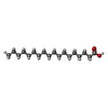 ChemComp-STE: |
-Macromolecule #7: MYRISTIC ACID
| Macromolecule | Name: MYRISTIC ACID / type: ligand / ID: 7 / Number of copies: 1 / Formula: MYR |
|---|---|
| Molecular weight | Theoretical: 228.371 Da |
| Chemical component information |  ChemComp-MYR: |
-Macromolecule #8: 2-acetamido-2-deoxy-beta-D-glucopyranose
| Macromolecule | Name: 2-acetamido-2-deoxy-beta-D-glucopyranose / type: ligand / ID: 8 / Number of copies: 2 / Formula: NAG |
|---|---|
| Molecular weight | Theoretical: 221.208 Da |
| Chemical component information |  ChemComp-NAG: |
-Experimental details
-Structure determination
| Method | cryo EM |
|---|---|
 Processing Processing | single particle reconstruction |
| Aggregation state | particle |
-Sample preparation
| Buffer | pH: 7.4 |
|---|---|
| Grid | Model: EMS Lacey Carbon / Material: COPPER / Mesh: 200 / Support film - Material: CARBON / Support film - topology: LACEY / Pretreatment - Type: GLOW DISCHARGE / Pretreatment - Time: 60 sec. / Pretreatment - Atmosphere: AIR |
| Vitrification | Cryogen name: ETHANE / Chamber humidity: 100 % / Chamber temperature: 273.0 K / Instrument: FEI VITROBOT MARK IV |
- Electron microscopy
| Microscope | JEOL CRYO ARM 300 |
|---|---|
| Image recording | Film or detector model: GATAN K3 BIOCONTINUUM (6k x 4k) / Number grids imaged: 1 / Number real images: 5004 / Average exposure time: 2.7 sec. / Average electron dose: 40.0 e/Å2 |
| Electron beam | Acceleration voltage: 300 kV / Electron source:  FIELD EMISSION GUN FIELD EMISSION GUN |
| Electron optics | Illumination mode: FLOOD BEAM / Imaging mode: BRIGHT FIELD / Cs: 2.7 mm / Nominal defocus max: 2.5 µm / Nominal defocus min: 0.5 µm |
| Sample stage | Specimen holder model: JEOL CRYOSPECPORTER / Cooling holder cryogen: NITROGEN |

