Movie
Movie Controller

[English] 日本語
 Yorodumi
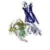
- EMDB-64907: Cryo-EM structure of human Neurotensin Receptor 1 (hNTSR1)-Gq (de... -
+ Open data

- Basic information
| Entry |  | ||||||||||||||||||||||||
|---|---|---|---|---|---|---|---|---|---|---|---|---|---|---|---|---|---|---|---|---|---|---|---|---|---|
| Title | Cryo-EM structure of human Neurotensin Receptor 1 (hNTSR1)-Gq (delipidated) complex in nucleotide-free C state | ||||||||||||||||||||||||
 Map data Map data | |||||||||||||||||||||||||
 Sample Sample |
| ||||||||||||||||||||||||
 Keywords Keywords | GPCR / NTSR1 / neurotensin / G-protein / membrane protein | ||||||||||||||||||||||||
| Function / homology |  Function and homology information Function and homology informationFatty Acids bound to GPR40 (FFAR1) regulate insulin secretion / G protein-coupled neurotensin receptor activity / Acetylcholine regulates insulin secretion / inositol phosphate catabolic process / sensory perception of itch / symmetric synapse / positive regulation of locomotion / phospholipase C-activating G protein-coupled glutamate receptor signaling pathway / regulation of inositol trisphosphate biosynthetic process / phospholipase C-activating serotonin receptor signaling pathway ...Fatty Acids bound to GPR40 (FFAR1) regulate insulin secretion / G protein-coupled neurotensin receptor activity / Acetylcholine regulates insulin secretion / inositol phosphate catabolic process / sensory perception of itch / symmetric synapse / positive regulation of locomotion / phospholipase C-activating G protein-coupled glutamate receptor signaling pathway / regulation of inositol trisphosphate biosynthetic process / phospholipase C-activating serotonin receptor signaling pathway / regulation of platelet activation / PLC beta mediated events / positive regulation of gamma-aminobutyric acid secretion / entrainment of circadian clock / D-aspartate import across plasma membrane / positive regulation of arachidonate secretion / vocalization behavior / L-glutamate import across plasma membrane / regulation of behavioral fear response / cAMP biosynthetic process / regulation of respiratory gaseous exchange / negative regulation of systemic arterial blood pressure / positive regulation of inhibitory postsynaptic potential / regulation of canonical Wnt signaling pathway / negative regulation of release of sequestered calcium ion into cytosol / glutamate receptor signaling pathway / response to food / positive regulation of glutamate secretion / regulation of membrane depolarization / response to lipid / temperature homeostasis / positive regulation of inositol phosphate biosynthetic process / response to stress / detection of temperature stimulus involved in sensory perception of pain / phototransduction, visible light / photoreceptor outer segment / postsynaptic cytosol / conditioned place preference / hormone-mediated signaling pathway / cellular response to acidic pH / Turbulent (oscillatory, disturbed) flow shear stress activates signaling by PIEZO1 and integrins in endothelial cells / positive regulation of release of sequestered calcium ion into cytosol / mast cell degranulation / GTPase activator activity / Peptide ligand-binding receptors / dendritic shaft / neuropeptide signaling pathway / response to prostaglandin E / G protein-coupled receptor binding / G protein-coupled receptor activity / cytoplasmic side of plasma membrane / G-protein beta/gamma-subunit complex binding / blood coagulation / Olfactory Signaling Pathway / terminal bouton / Activation of the phototransduction cascade / G protein-coupled acetylcholine receptor signaling pathway / G beta:gamma signalling through PLC beta / Presynaptic function of Kainate receptors / Thromboxane signalling through TP receptor / Activation of G protein gated Potassium channels / Inhibition of voltage gated Ca2+ channels via Gbeta/gamma subunits / G-protein activation / Glucagon signaling in metabolic regulation / G beta:gamma signalling through CDC42 / Prostacyclin signalling through prostacyclin receptor / Synthesis, secretion, and inactivation of Glucagon-like Peptide-1 (GLP-1) / G beta:gamma signalling through BTK / photoreceptor disc membrane / ADP signalling through P2Y purinoceptor 12 / Glucagon-type ligand receptors / Sensory perception of sweet, bitter, and umami (glutamate) taste / Adrenaline,noradrenaline inhibits insulin secretion / Vasopressin regulates renal water homeostasis via Aquaporins / Glucagon-like Peptide-1 (GLP1) regulates insulin secretion / G alpha (z) signalling events / cellular response to catecholamine stimulus / ADP signalling through P2Y purinoceptor 1 / G beta:gamma signalling through PI3Kgamma / ADORA2B mediated anti-inflammatory cytokines production / adenylate cyclase-activating dopamine receptor signaling pathway / Cooperation of PDCL (PhLP1) and TRiC/CCT in G-protein beta folding / GPER1 signaling / cellular response to prostaglandin E stimulus / heterotrimeric G-protein complex / Inactivation, recovery and regulation of the phototransduction cascade / G alpha (12/13) signalling events / G-protein beta-subunit binding / extracellular vesicle / sensory perception of taste / Thrombin signalling through proteinase activated receptors (PARs) / signaling receptor complex adaptor activity / adenylate cyclase-activating G protein-coupled receptor signaling pathway / retina development in camera-type eye / fibroblast proliferation / nuclear membrane / GTPase binding / G protein activity / Ca2+ pathway / chemical synaptic transmission Similarity search - Function | ||||||||||||||||||||||||
| Biological species |  Homo sapiens (human) / synthetic construct (others) / Homo sapiens (human) / synthetic construct (others) /  | ||||||||||||||||||||||||
| Method | single particle reconstruction / Resolution: 2.24 Å | ||||||||||||||||||||||||
 Authors Authors | Matsui TE / Kobayashi K / Fukuda M / Kawakami K / Yamashita K / Kato HE | ||||||||||||||||||||||||
| Funding support |  Japan, 7 items Japan, 7 items
| ||||||||||||||||||||||||
 Citation Citation | Journal: Nature / Year: 2026 Title: The dynamic basis of G-protein recognition and activation by a GPCR. Authors: Kazuhiro Kobayashi / Kouki Kawakami / Toshiki E Matsui / Shun Yokoi / Masahiro Fukuda / Tomohiro J Narita / Hiroki Arai / Mai Tambo / Takashi Sumikama / Manae Tatsumi / Keitaro Yamashita / ...Authors: Kazuhiro Kobayashi / Kouki Kawakami / Toshiki E Matsui / Shun Yokoi / Masahiro Fukuda / Tomohiro J Narita / Hiroki Arai / Mai Tambo / Takashi Sumikama / Manae Tatsumi / Keitaro Yamashita / Junki Koyanagi / Mai Kugawa / Hisako Ikeda / Ayumi Sumino / Ayori Mitsutake / Brian K Kobilka / Asuka Inoue / Hideaki E Kato /  Abstract: G-protein-coupled receptor (GPCR) signalling occurs through heterotrimeric G proteins, whose selective activation leads to distinct cellular outcomes. Although more than 200 GPCR-G protein complex ...G-protein-coupled receptor (GPCR) signalling occurs through heterotrimeric G proteins, whose selective activation leads to distinct cellular outcomes. Although more than 200 GPCR-G protein complex structures have been determined, these static snapshots provide limited insight into the dynamics of G-protein association and dissociation. Here we present cryo-electron microscopy structures of human neurotensin receptor type 1 (NTSR1) with minimally modified G and G, showing how the receptor's intracellular surface dynamically rearranges to accommodate each G-protein subtype. Furthermore, time-resolved cryo-electron microscopy analyses of NTSR1-G visualized G-protein dissociation processes on GDP/GTP binding. Characterization of more than 20 intermediates, complemented by mutational and computational analyses, identifies four key mechanistic features. First, GDP/GTP induces G release from both canonical and non-canonical active conformations with distinct kinetics. Second, NTSR1 uses common intracellular rearrangements to recognize different G-protein subtypes and to promote activation of a single subtype. Third, separation from Gβγ involves stepwise remodelling of the Gα switches I-III. Finally, G dissociates from the receptor through a pathway that is distinct from that of G, and the canonical and non-canonical NTSR1-G complexes further diverge in their dissociation trajectories. These findings provide a comprehensive framework for understanding GPCR signalling dynamics and guiding signal-targeted therapeutic development. | ||||||||||||||||||||||||
| History |
|
- Structure visualization
| Supplemental images |
|---|

- Downloads & links
-EMDB archive
| Map data | emd_64907.map.gz | 15.5 MB | EMDB map data format | |
|---|---|---|---|---|
| Header (meta data) | emd-64907-v30.xmlemd-64907.xml | 29.1 KB 29.1 KB | Display Display | EMDB header |
| FSC (resolution estimation) | emd_64907_fsc.xml | 11.7 KB | Display | FSC data file |
| Images |  emd_64907.png emd_64907.png | 118 KB | ||
| Masks | emd_64907_msk_1.map | 16.8 MB | Mask map | |
| Filedesc metadata | emd-64907.cif.gz | 7.4 KB | ||
| Others | emd_64907_additional_1.map.gzemd_64907_half_map_1.map.gzemd_64907_half_map_2.map.gz | 3.3 MB 15.5 MB 15.5 MB | ||
| Archive directory |  http://ftp.pdbj.org/pub/emdb/structures/EMD-64907ftp://ftp.pdbj.org/pub/emdb/structures/EMD-64907 http://ftp.pdbj.org/pub/emdb/structures/EMD-64907ftp://ftp.pdbj.org/pub/emdb/structures/EMD-64907 | HTTPS FTP |
-Related structure data
| Related structure data |  9vawMC  20zcC  20zdC  20zgC  20zhC  20ziC  20zjC  20zkC  20zlC  9vatC  9vauC  9vavC  9vaxC  9vayC  9vazC  9vb0C  9vb1C  9vb2C  9vb3C  9vb4C  9vb5C  9vb6C  9vb7C  9vbaC M: atomic model generated by this map C: citing same article ( |
|---|---|
| Similar structure data |


-Links
| EMDB pages | EMDB (EBI/PDBe) / EMDataResource |
|---|---|
| Related items in Molecule of the Month |


-Map
| File | Download / File: emd_64907.map.gz / Format: CCP4 / Size: 16.8 MB / Type: IMAGE STORED AS FLOATING POINT NUMBER (4 BYTES) | ||||||||||||||||||||||||||||||||||||
|---|---|---|---|---|---|---|---|---|---|---|---|---|---|---|---|---|---|---|---|---|---|---|---|---|---|---|---|---|---|---|---|---|---|---|---|---|---|
| Projections & slices | Image control
Images are generated by Spider. | ||||||||||||||||||||||||||||||||||||
| Voxel size | X=Y=Z: 1.06 Å | ||||||||||||||||||||||||||||||||||||
| Density |
| ||||||||||||||||||||||||||||||||||||
| Symmetry | Space group: 1 | ||||||||||||||||||||||||||||||||||||
| Details | EMDB XML:
|
 Z (Sec.)
 Y (Row.)
 X (Col.)


-Supplemental data
-Mask #1
| File | emd_64907_msk_1.map | ||||||||||||
|---|---|---|---|---|---|---|---|---|---|---|---|---|---|
| Projections & Slices |
| ||||||||||||
| Density Histograms |


-Additional map: #1
| File | emd_64907_additional_1.map | ||||||||||||
|---|---|---|---|---|---|---|---|---|---|---|---|---|---|
| Projections & Slices |
| ||||||||||||
| Density Histograms |


-Half map: #2
| File | emd_64907_half_map_1.map | ||||||||||||
|---|---|---|---|---|---|---|---|---|---|---|---|---|---|
| Projections & Slices |
| ||||||||||||
| Density Histograms |


-Half map: #1
| File | emd_64907_half_map_2.map | ||||||||||||
|---|---|---|---|---|---|---|---|---|---|---|---|---|---|
| Projections & Slices |
| ||||||||||||
| Density Histograms |


- Sample components
+Entire : JMV449-bound human Neurotensin Receptor 1 (hNTSR1)-GqiN18 complex...
+Supramolecule #1: JMV449-bound human Neurotensin Receptor 1 (hNTSR1)-GqiN18 complex...
+Supramolecule #2: JMV449-bound human neurotensin receptor type 1
+Supramolecule #3: heterotrimeric GqiN18 protein with delipidation mutations
+Supramolecule #4: scFv16
+Macromolecule #1: Neurotensin receptor type 1

 Spodoptera frugiperda (fall armyworm)
+Macromolecule #2: JMV449
+Macromolecule #3: Guanine nucleotide-binding protein G(q) subunit alpha
+Macromolecule #4: Guanine nucleotide-binding protein G(I)/G(S)/G(T) subunit beta-1
+Macromolecule #5: Guanine nucleotide-binding protein G(I)/G(S)/G(O) subunit gamma-2
+Macromolecule #6: scFv16
 Trichoplusia ni ascovirus 2a
+Macromolecule #7: water

-Experimental details
-Structure determination
 Processing Processing | single particle reconstruction |
|---|---|
| Aggregation state | particle |
-Sample preparation
| Buffer | pH: 7.5 |
|---|---|
| Grid | Model: Quantifoil R1.2/1.3 / Material: GOLD / Mesh: 300 / Support film - Material: GOLD / Support film - topology: HOLEY / Pretreatment - Type: GLOW DISCHARGE |
- Electron microscopy
| Microscope | TFS KRIOS |
|---|---|
| Image recording | Film or detector model: GATAN K3 BIOQUANTUM (6k x 4k) / Average electron dose: 55.0 e/Å2 |
| Electron beam | Acceleration voltage: 300 kV / Electron source:  FIELD EMISSION GUN FIELD EMISSION GUN |
| Electron optics | Illumination mode: OTHER / Imaging mode: BRIGHT FIELD / Cs: 2.7 mm / Nominal defocus max: 1.6 µm / Nominal defocus min: 0.8 µm |
| Sample stage | Cooling holder cryogen: NITROGEN |
| Experimental equipment |  Model: Titan Krios / Image courtesy: FEI Company |