 Movie
Movie Controller


+ Open data

- Basic information
| Entry | Database: EMDB / ID: EMD-6482 | |||||||||
|---|---|---|---|---|---|---|---|---|---|---|
| Title | Cryo-electron microscopy of alpha Synuclein amyloid fibrils | |||||||||
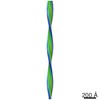
 Map data Map data | Extruded 2D reconstruction of in vitro assembled alpha Synuclein amyoid fibrils | |||||||||
 Sample Sample |
| |||||||||
| Function / homology |  Function and homology information Function and homology informationprotein binding / negative regulation of mitochondrial electron transport, NADH to ubiquinone / neutral lipid metabolic process / regulation of acyl-CoA biosynthetic process / negative regulation of dopamine uptake involved in synaptic transmission / negative regulation of norepinephrine uptake / response to desipramine / positive regulation of SNARE complex assembly / positive regulation of hydrogen peroxide catabolic process / supramolecular fiber ...protein binding / negative regulation of mitochondrial electron transport, NADH to ubiquinone / neutral lipid metabolic process / regulation of acyl-CoA biosynthetic process / negative regulation of dopamine uptake involved in synaptic transmission / negative regulation of norepinephrine uptake / response to desipramine / positive regulation of SNARE complex assembly / positive regulation of hydrogen peroxide catabolic process / supramolecular fiber / regulation of synaptic vesicle recycling / negative regulation of chaperone-mediated autophagy / mitochondrial membrane organization / regulation of reactive oxygen species biosynthetic process / positive regulation of protein localization to cell periphery / negative regulation of exocytosis / regulation of glutamate secretion / dopamine biosynthetic process / regulation of macrophage activation / positive regulation of neurotransmitter secretion / response to iron(II) ion / negative regulation of dopamine metabolic process / negative regulation of platelet-derived growth factor receptor signaling pathway / SNARE complex assembly / negative regulation of thrombin-activated receptor signaling pathway / Lewy body / regulation of locomotion / negative regulation of microtubule polymerization / synaptic vesicle priming / regulation of norepinephrine uptake / transporter regulator activity / protein kinase inhibitor activity / positive regulation of inositol phosphate biosynthetic process / synaptic vesicle transport / dopamine uptake involved in synaptic transmission / regulation of dopamine secretion / positive regulation of receptor recycling / cuprous ion binding / positive regulation of exocytosis / nuclear outer membrane / mitochondrial ATP synthesis coupled electron transport / dynein complex binding / synaptic vesicle exocytosis / response to magnesium ion / positive regulation of endocytosis / negative regulation of serotonin uptake / response to type II interferon / cysteine-type endopeptidase inhibitor activity / kinesin binding / regulation of presynapse assembly / synaptic vesicle endocytosis / alpha-tubulin binding / beta-tubulin binding / phospholipase binding / phospholipid metabolic process / supramolecular fiber organization / behavioral response to cocaine / cellular response to fibroblast growth factor stimulus / cellular response to epinephrine stimulus / inclusion body / Hsp70 protein binding / enzyme inhibitor activity / response to interleukin-1 / axon terminus / cellular response to copper ion / fatty acid binding / regulation of microtubule cytoskeleton organization / positive regulation of release of sequestered calcium ion into cytosol / SNARE binding / adult locomotory behavior / glutathione metabolic process / protein tetramerization / protein sequestering activity / phosphoprotein binding / excitatory postsynaptic potential / tubulin binding / microglial cell activation / ferrous iron binding / fatty acid metabolic process / phospholipid binding / PKR-mediated signaling / synapse organization / receptor internalization / regulation of long-term neuronal synaptic plasticity / protein destabilization / tau protein binding / enzyme activator activity / positive regulation of inflammatory response / terminal bouton / long-term synaptic potentiation / actin cytoskeleton / synaptic vesicle membrane / growth cone / actin binding / cellular response to oxidative stress / neuron apoptotic process / histone binding / cell cortex / response to lipopolysaccharide / microtubule binding Similarity search - Function | |||||||||
| Biological species |  Homo sapiens (human) Homo sapiens (human) | |||||||||
| Method | helical reconstruction / cryo EM / Resolution: 40.0 Å | |||||||||
 Authors Authors | Dearborn AD / Wall JS / Cheng N / Heymann JB / Kajava AV / Varkey J / Langen R / Steven AC | |||||||||
 Citation Citation | Journal: J Biol Chem / Year: 2016 Title: α-Synuclein Amyloid Fibrils with Two Entwined, Asymmetrically Associated Protofibrils. Authors: Altaira D Dearborn / Joseph S Wall / Naiqian Cheng / J Bernard Heymann / Andrey V Kajava / Jobin Varkey / Ralf Langen / Alasdair C Steven /    Abstract: Parkinson disease and other progressive neurodegenerative conditions are characterized by the intracerebral presence of Lewy bodies, containing amyloid fibrils of α-synuclein. We used cryo-electron ...Parkinson disease and other progressive neurodegenerative conditions are characterized by the intracerebral presence of Lewy bodies, containing amyloid fibrils of α-synuclein. We used cryo-electron microscopy and scanning transmission electron microscopy (STEM) to study in vitro-assembled fibrils. These fibrils are highly polymorphic. Focusing on twisting fibrils with an inter-crossover spacing of 77 nm, our reconstructions showed them to consist of paired protofibrils. STEM mass per length data gave one subunit per 0.47 nm axial rise per protofibril, consistent with a superpleated β-structure. The STEM images show two thread-like densities running along each of these fibrils, which we interpret as ladders of metal ions. These threads confirmed the two-protofibril architecture of the 77-nm twisting fibrils and allowed us to identify this morphotype in STEM micrographs. Some other, but not all, fibril morphotypes also exhibit dense threads, implying that they also present a putative metal binding site. We propose a molecular model for the protofibril and suggest that polymorphic variant fibrils have different numbers of protofibrils that are associated differently. | |||||||||
| History |
|
- Structure visualization
| Movie |
Movie viewer |
|---|---|
| Structure viewer | EM map: SurfViewMolmilJmol/JSmol |
| Supplemental images |

- Downloads & links
-EMDB archive
| Map data | emd_6482.map.gz | 13.8 MB | EMDB map data format | |
|---|---|---|---|---|
| Header (meta data) | emd-6482-v30.xmlemd-6482.xml | 11.1 KB 11.1 KB | Display Display | EMDB header |
| Images | 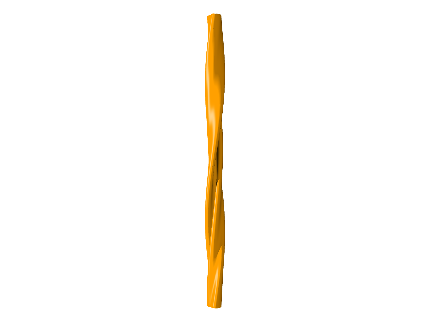 emd_6482.png emd_6482.png | 54.5 KB | ||
| Archive directory |  http://ftp.pdbj.org/pub/emdb/structures/EMD-6482ftp://ftp.pdbj.org/pub/emdb/structures/EMD-6482 http://ftp.pdbj.org/pub/emdb/structures/EMD-6482ftp://ftp.pdbj.org/pub/emdb/structures/EMD-6482 | HTTPS FTP |
-Related structure data
| Similar structure data |
|---|
-Links
| EMDB pages | EMDB (EBI/PDBe) / EMDataResource |
|---|---|
| Related items in Molecule of the Month |

-Map
| File | Download / File: emd_6482.map.gz / Format: CCP4 / Size: 14.4 MB / Type: IMAGE STORED AS FLOATING POINT NUMBER (4 BYTES) | ||||||||||||||||||||||||||||||||||||||||||||||||||||||||||||
|---|---|---|---|---|---|---|---|---|---|---|---|---|---|---|---|---|---|---|---|---|---|---|---|---|---|---|---|---|---|---|---|---|---|---|---|---|---|---|---|---|---|---|---|---|---|---|---|---|---|---|---|---|---|---|---|---|---|---|---|---|---|
| Annotation | Extruded 2D reconstruction of in vitro assembled alpha Synuclein amyoid fibrils | ||||||||||||||||||||||||||||||||||||||||||||||||||||||||||||
| Projections & slices | Image control
Images are generated by Spider. generated in cubic-lattice coordinate | ||||||||||||||||||||||||||||||||||||||||||||||||||||||||||||
| Voxel size | X=Y=Z: 2.54 Å | ||||||||||||||||||||||||||||||||||||||||||||||||||||||||||||
| Density |
| ||||||||||||||||||||||||||||||||||||||||||||||||||||||||||||
| Symmetry | Space group: 1 | ||||||||||||||||||||||||||||||||||||||||||||||||||||||||||||
| Details | EMDB XML:
CCP4 map header:
| ||||||||||||||||||||||||||||||||||||||||||||||||||||||||||||
 Z (Sec.)
 Y (Row.)
 X (Col.)


-Supplemental data
- Sample components
-Entire : In vitro assembled, recombinant amyloid fibrils of full-length hu...
| Entire | Name: In vitro assembled, recombinant amyloid fibrils of full-length human alpha Synuclein |
|---|---|
| Components |
|
-Supramolecule #1000: In vitro assembled, recombinant amyloid fibrils of full-length hu...
| Supramolecule | Name: In vitro assembled, recombinant amyloid fibrils of full-length human alpha Synuclein type: sample / ID: 1000 / Details: The sample was morphologically heterogeneous. / Oligomeric state: dimeric asymmetric unit / Number unique components: 2 |
|---|---|
| Molecular weight | Method: Dark-field scanning transmission electron microscopy 59.1 MDa/micron compared to 61.5 MDa/micron theoretical weight |
-Macromolecule #1: alpha Synuclein
| Macromolecule | Name: alpha Synuclein / type: protein_or_peptide / ID: 1 / Name.synonym: NACP, PARK1 / Oligomeric state: Amyloid / Recombinant expression: Yes |
|---|---|
| Source (natural) | Organism: Homo sapiens (human) / synonym: Human / Tissue: Brain / Cell: Neuron / Location in cell: Lewy Body |
| Recombinant expression | Organism:  |
| Sequence | UniProtKB: Alpha-synuclein GO: magnesium ion binding, fatty acid binding, copper ion binding, calcium ion binding, protein binding InterPro: Alpha-synuclein |
-Experimental details
-Structure determination
| Method | cryo EM |
|---|---|
 Processing Processing | helical reconstruction |
| Aggregation state | filament |
-Sample preparation
| Concentration | 4 mg/mL |
|---|---|
| Buffer | pH: 7.4 / Details: 10 mM HEPES, 100 mM NaCl, 0.1% NaN3 |
| Grid | Details: R1.2/1.3 400 mesh copper Quantifoil grid, glow-discharged in argon/oxygen |
| Vitrification | Cryogen name: ETHANE / Chamber humidity: 90 % / Chamber temperature: 90 K / Instrument: LEICA KF80 / Method: Manually blotted before plunging |
- Electron microscopy
| Microscope | FEI/PHILIPS CM200FEG |
|---|---|
| Date | Jun 5, 2012 |
| Image recording | Category: FILM / Film or detector model: KODAK SO-163 FILM / Digitization - Scanner: NIKON SUPER COOLSCAN 9000 / Digitization - Sampling interval: 6.35 µm / Number real images: 110 / Average electron dose: 15 e/Å2 / Bits/pixel: 16 |
| Electron beam | Acceleration voltage: 120 kV / Electron source:  FIELD EMISSION GUN FIELD EMISSION GUN |
| Electron optics | Calibrated magnification: 51840 / Illumination mode: FLOOD BEAM / Imaging mode: BRIGHT FIELD / Cs: 2.0 mm / Nominal defocus max: 3.5 µm / Nominal defocus min: 2.0 µm / Nominal magnification: 50000 |
| Sample stage | Specimen holder model: GATAN LIQUID NITROGEN |
-Image processing
| Details | The 2D reconstructions were made and aligned using bhelcross. The helical parameters were determined per fibril in real space and imposed on the average using bhelcross. |
|---|---|
| Final reconstruction | Applied symmetry - Helical parameters - Δz: 4.7 Å Applied symmetry - Helical parameters - Δ&Phi: 1.1 ° Applied symmetry - Helical parameters - Axial symmetry: C1 (asymmetric) Resolution.type: BY AUTHOR / Resolution: 40.0 Å / Resolution method: OTHER / Software - Name: Bsoft Details: Extruded from a 2D reconstruction based upon helical parameters |
| CTF correction | Details: phase-flipped micrograph |
