Movie
Movie Controller


+ Open data

- Basic information
| Entry |  | |||||||||
|---|---|---|---|---|---|---|---|---|---|---|
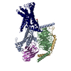
| Title | GPR133-Gain-miniG13 complex | |||||||||
 Map data Map data | ||||||||||
 Sample Sample |
| |||||||||
 Keywords Keywords | GPCR / G12/G13 / Complex / Stachel / MEMBRANE PROTEIN | |||||||||
| Function / homology |  Function and homology information Function and homology informationD5 dopamine receptor binding / regulation of fibroblast migration / Rho-activating G protein-coupled receptor signaling pathway / regulation of small GTPase mediated signal transduction / NRAGE signals death through JNK / branching involved in blood vessel morphogenesis / CDC42 GTPase cycle / regulation of skeletal muscle contraction / regulation of postsynapse assembly / Rho protein signal transduction ...D5 dopamine receptor binding / regulation of fibroblast migration / Rho-activating G protein-coupled receptor signaling pathway / regulation of small GTPase mediated signal transduction / NRAGE signals death through JNK / branching involved in blood vessel morphogenesis / CDC42 GTPase cycle / regulation of skeletal muscle contraction / regulation of postsynapse assembly / Rho protein signal transduction / RAC1 GTPase cycle / guanyl-nucleotide exchange factor activity / brush border membrane / platelet activation / regulation of blood pressure / G protein-coupled receptor activity / G-protein beta/gamma-subunit complex binding / adenylate cyclase-modulating G protein-coupled receptor signaling pathway / Olfactory Signaling Pathway / Activation of the phototransduction cascade / G protein-coupled acetylcholine receptor signaling pathway / G beta:gamma signalling through PLC beta / Presynaptic function of Kainate receptors / Thromboxane signalling through TP receptor / Activation of G protein gated Potassium channels / Inhibition of voltage gated Ca2+ channels via Gbeta/gamma subunits / G-protein activation / Glucagon signaling in metabolic regulation / Prostacyclin signalling through prostacyclin receptor / G beta:gamma signalling through CDC42 / Synthesis, secretion, and inactivation of Glucagon-like Peptide-1 (GLP-1) / melanosome / G beta:gamma signalling through BTK / photoreceptor disc membrane / ADP signalling through P2Y purinoceptor 12 / Glucagon-type ligand receptors / Sensory perception of sweet, bitter, and umami (glutamate) taste / Adrenaline,noradrenaline inhibits insulin secretion / Vasopressin regulates renal water homeostasis via Aquaporins / Glucagon-like Peptide-1 (GLP1) regulates insulin secretion / G alpha (z) signalling events / cellular response to catecholamine stimulus / ADP signalling through P2Y purinoceptor 1 / regulation of cell shape / ADORA2B mediated anti-inflammatory cytokines production / G beta:gamma signalling through PI3Kgamma / adenylate cyclase-activating dopamine receptor signaling pathway / Cooperation of PDCL (PhLP1) and TRiC/CCT in G-protein beta folding / GPER1 signaling / cellular response to prostaglandin E stimulus / heterotrimeric G-protein complex / G alpha (12/13) signalling events / G-protein beta-subunit binding / Inactivation, recovery and regulation of the phototransduction cascade / extracellular vesicle / sensory perception of taste / Thrombin signalling through proteinase activated receptors (PARs) / adenylate cyclase-activating G protein-coupled receptor signaling pathway / signaling receptor complex adaptor activity / retina development in camera-type eye / GTPase binding / fibroblast proliferation / G protein activity / Ca2+ pathway / High laminar flow shear stress activates signaling by PIEZO1 and PECAM1:CDH5:KDR in endothelial cells / G alpha (i) signalling events / G alpha (s) signalling events / phospholipase C-activating G protein-coupled receptor signaling pathway / G alpha (q) signalling events / in utero embryonic development / Ras protein signal transduction / cell differentiation / cell surface receptor signaling pathway / Extra-nuclear estrogen signaling / cell population proliferation / nuclear speck / postsynapse / G protein-coupled receptor signaling pathway / lysosomal membrane / focal adhesion / GTPase activity / synapse / GTP binding / protein-containing complex binding / signal transduction / extracellular exosome / nucleoplasm / membrane / metal ion binding / nucleus / plasma membrane / cytoplasm / cytosol Similarity search - Function | |||||||||
| Biological species |  Homo sapiens (human) Homo sapiens (human) | |||||||||
| Method | single particle reconstruction / cryo EM / Resolution: 3.51 Å | |||||||||
 Authors Authors | Xi Y / Pu X / Ping Y | |||||||||
| Funding support |  China, 1 items China, 1 items
| |||||||||
 Citation Citation | Journal: Biochem Biophys Res Commun / Year: 2025 Title: Cryo-EM structural elucidation and molecular mechanism of the GPR133-G13 signaling complex. Authors: Xuanyu Pu / Yue-Tong Xi / Meng-Xin Wang / Daolai Zhang / Yu-Qi Ping / Peng Xiao / Jin-Peng Sun / Abstract: GPR133 is an adhesion-class G protein-coupled receptor (GPCR) that has recently been de-orphanized. Its functions are complex and multifaceted. While GPR133 is primarily recognized for coupling with ...GPR133 is an adhesion-class G protein-coupled receptor (GPCR) that has recently been de-orphanized. Its functions are complex and multifaceted. While GPR133 is primarily recognized for coupling with the Gs subunit to mediate elevated intracellular cAMP levels, its potential engagement with alternative signaling pathways remains poorly characterized. In our experiments, we demonstrated that GPR133 exhibits constitutive self-activation via its Stachel sequence as an adhesion GPCR, enabling activation of downstream G13 signaling. We reconstituted the GPR133-GAIN-miniGα13 complex in vitro and resolved its cryo-electron microscopy structure at a resolution of 3.51 Å. Detailed structural comparisons between the GPR133-GAIN-miniGα13 complex and the previously resolved GPR133-CTF-Gs structure highlighted both conserved and different features. These findings provide critical insights into the signal transduction mechanisms of GPR133 and lay a foundation for targeted therapeutic strategies. | |||||||||
| History |
|
- Structure visualization
| Supplemental images |
|---|

- Downloads & links
-EMDB archive
| Map data | emd_64672.map.gz | 52.8 MB | EMDB map data format | |
|---|---|---|---|---|
| Header (meta data) | emd-64672-v30.xmlemd-64672.xml | 18.1 KB 18.1 KB | Display Display | EMDB header |
| Images |  emd_64672.png emd_64672.png | 34.9 KB | ||
| Filedesc metadata | emd-64672.cif.gz | 6.2 KB | ||
| Others | emd_64672_half_map_1.map.gzemd_64672_half_map_2.map.gz | 55.4 MB 55.4 MB | ||
| Archive directory |  http://ftp.pdbj.org/pub/emdb/structures/EMD-64672ftp://ftp.pdbj.org/pub/emdb/structures/EMD-64672 http://ftp.pdbj.org/pub/emdb/structures/EMD-64672ftp://ftp.pdbj.org/pub/emdb/structures/EMD-64672 | HTTPS FTP |
-Related structure data
| Related structure data |  9v0uMC M: atomic model generated by this map C: citing same article ( |
|---|---|
| Similar structure data |
-Links
| EMDB pages | EMDB (EBI/PDBe) / EMDataResource |
|---|---|
| Related items in Molecule of the Month |


-Map
| File | Download / File: emd_64672.map.gz / Format: CCP4 / Size: 59.6 MB / Type: IMAGE STORED AS FLOATING POINT NUMBER (4 BYTES) | ||||||||||||||||||||||||||||||||||||
|---|---|---|---|---|---|---|---|---|---|---|---|---|---|---|---|---|---|---|---|---|---|---|---|---|---|---|---|---|---|---|---|---|---|---|---|---|---|
| Projections & slices | Image control
Images are generated by Spider. | ||||||||||||||||||||||||||||||||||||
| Voxel size | X=Y=Z: 0.93 Å | ||||||||||||||||||||||||||||||||||||
| Density |
| ||||||||||||||||||||||||||||||||||||
| Symmetry | Space group: 1 | ||||||||||||||||||||||||||||||||||||
| Details | EMDB XML:
|
 Z (Sec.)
 Y (Row.)
 X (Col.)


-Supplemental data
-Half map: #2
| File | emd_64672_half_map_1.map | ||||||||||||
|---|---|---|---|---|---|---|---|---|---|---|---|---|---|
| Projections & Slices |
| ||||||||||||
| Density Histograms |


-Half map: #1
| File | emd_64672_half_map_2.map | ||||||||||||
|---|---|---|---|---|---|---|---|---|---|---|---|---|---|
| Projections & Slices |
| ||||||||||||
| Density Histograms |


- Sample components
-Entire : GPR133-Gain-miniG13-Gbeta1-Ggamma2 complex
| Entire | Name: GPR133-Gain-miniG13-Gbeta1-Ggamma2 complex |
|---|---|
| Components |
|
-Supramolecule #1: GPR133-Gain-miniG13-Gbeta1-Ggamma2 complex
| Supramolecule | Name: GPR133-Gain-miniG13-Gbeta1-Ggamma2 complex / type: complex / ID: 1 / Parent: 0 / Macromolecule list: all |
|---|---|
| Source (natural) | Organism: Homo sapiens (human) |
-Macromolecule #1: Adhesion G-protein coupled receptor D1
| Macromolecule | Name: Adhesion G-protein coupled receptor D1 / type: protein_or_peptide / ID: 1 / Number of copies: 1 / Enantiomer: LEVO |
|---|---|
| Source (natural) | Organism: Homo sapiens (human) |
| Molecular weight | Theoretical: 65.688828 KDa |
| Recombinant expression | Organism:   Spodoptera frugiperda (fall armyworm) Spodoptera frugiperda (fall armyworm) |
| Sequence | String: HPIITNLTEE RKTFQSPGVI LSYLQNVSLS LPSKSLSEQT ALNLTKTFLK AVGEILLLPG WIALSEDSAV VLSLIDTIDT VMGHVSSNL HGSTPQVTVE GSSAMAEFSV AKILPKTVNS SHYRFPAHGQ SFIQIPHEAF HRHAWSTVVG LLYHSMHYYL N NIWPAHTK ...String: HPIITNLTEE RKTFQSPGVI LSYLQNVSLS LPSKSLSEQT ALNLTKTFLK AVGEILLLPG WIALSEDSAV VLSLIDTIDT VMGHVSSNL HGSTPQVTVE GSSAMAEFSV AKILPKTVNS SHYRFPAHGQ SFIQIPHEAF HRHAWSTVVG LLYHSMHYYL N NIWPAHTK IAEAMHHQDC LLFATSHLIS LEVSPPPTLS QNLSGSPLIT VHLKHRLTRK QHSEATNSSN RVFVYCAFLD FS SGEGVWS NHGCALTRGN LTYSVCRCTH LTNFAILMQV VPLELARGHQ VALSSISYVG CSLSVLCLVA TLVTFAVLSS VST IRNQRY HIHANLSFAV LVAQVLLLIS FRLEPGTTPC QVMAVLLHYF FLSAFAWMLV EGLHLYSMVI KVFGSEDSKH RYYY GMGWG FPLLICIISL SFAMDSYGTS NNCWLSLASG AIWAFVAPAL FVIVVNIGIL IAVTRVISQI SADNYKIHGD PSAFK LTAK AVAVLLPILG TSWVFGVLAV NGCAVVFQYM FATLNSLQGL FIFLFHCLLN SEVRAAFKHK TKVWSLTSSS ARTSNA KPF HSDLMNGTRP GMASTKLSPW DKSSHSAHRV DLSAV UniProtKB: Adhesion G-protein coupled receptor D1 |
-Macromolecule #2: Guanine nucleotide-binding protein subunit alpha-13,Isoform 2 of ...
| Macromolecule | Name: Guanine nucleotide-binding protein subunit alpha-13,Isoform 2 of Guanine nucleotide-binding protein subunit alpha-13 type: protein_or_peptide / ID: 2 / Number of copies: 1 / Enantiomer: LEVO |
|---|---|
| Source (natural) | Organism: Homo sapiens (human) |
| Molecular weight | Theoretical: 26.705596 KDa |
| Recombinant expression | Organism: Spodoptera frugiperda (fall armyworm) |
| Sequence | String: MMGSTVSAED KAAAERSKEI DKCLSREKTY VKRLVKILLL GADNSGKSTF LKQMRIIHGG SGGSGGTKGI HEYDFEIKNV PFKMVDVGG QRSERKRWFE CFDSVTSILF LVDSSDFNRL TESLNDFETI VNNRVFSNVS IILFLNKTDL LEEKVQIVSI K DYFLEFEG ...String: MMGSTVSAED KAAAERSKEI DKCLSREKTY VKRLVKILLL GADNSGKSTF LKQMRIIHGG SGGSGGTKGI HEYDFEIKNV PFKMVDVGG QRSERKRWFE CFDSVTSILF LVDSSDFNRL TESLNDFETI VNNRVFSNVS IILFLNKTDL LEEKVQIVSI K DYFLEFEG DPHCLRDVQK FLVECFRNKR RDQQQKPLYH HFTTAINTEN ARLIFRDVKD TILHDNLKQL MLQ UniProtKB: Guanine nucleotide-binding protein subunit alpha-13, Guanine nucleotide-binding protein subunit alpha-13, Guanine nucleotide-binding protein subunit alpha-13 |
-Macromolecule #3: Guanine nucleotide-binding protein G(I)/G(S)/G(O) subunit gamma-2
| Macromolecule | Name: Guanine nucleotide-binding protein G(I)/G(S)/G(O) subunit gamma-2 type: protein_or_peptide / ID: 3 / Number of copies: 1 / Enantiomer: LEVO |
|---|---|
| Source (natural) | Organism: Homo sapiens (human) |
| Molecular weight | Theoretical: 7.861143 KDa |
| Recombinant expression | Organism: Spodoptera frugiperda (fall armyworm) |
| Sequence | String: MASNNTASIA QARKLVEQLK MEANIDRIKV SKAAADLMAY CEAHAKEDPL LTPVPASENP FREKKFFCAI L UniProtKB: Guanine nucleotide-binding protein G(I)/G(S)/G(O) subunit gamma-2 |
-Macromolecule #4: Guanine nucleotide-binding protein G(I)/G(S)/G(T) subunit beta-1
| Macromolecule | Name: Guanine nucleotide-binding protein G(I)/G(S)/G(T) subunit beta-1 type: protein_or_peptide / ID: 4 / Number of copies: 1 / Enantiomer: LEVO |
|---|---|
| Source (natural) | Organism: Homo sapiens (human) |
| Molecular weight | Theoretical: 37.784301 KDa |
| Recombinant expression | Organism: Spodoptera frugiperda (fall armyworm) |
| Sequence | String: GSLLQSELDQ LRQEAEQLKN QIRDARKACA DATLSQITNN IDPVGRIQMR TRRTLRGHLA KIYAMHWGTD SRLLVSASQD GKLIIWDSY TTNKVHAIPL RSSWVMTCAY APSGNYVACG GLDNICSIYN LKTREGNVRV SRELAGHTGY LSCCRFLDDN Q IVTSSGDT ...String: GSLLQSELDQ LRQEAEQLKN QIRDARKACA DATLSQITNN IDPVGRIQMR TRRTLRGHLA KIYAMHWGTD SRLLVSASQD GKLIIWDSY TTNKVHAIPL RSSWVMTCAY APSGNYVACG GLDNICSIYN LKTREGNVRV SRELAGHTGY LSCCRFLDDN Q IVTSSGDT TCALWDIETG QQTTTFTGHT GDVMSLSLAP DTRLFVSGAC DASAKLWDVR EGMCRQTFTG HESDINAICF FP NGNAFAT GSDDATCRLF DLRADQELMT YSHDNIICGI TSVSFSKSGR LLLAGYDDFN CNVWDALKAD RAGVLAGHDN RVS CLGVTD DGMAVATGSW DSFLKIWN UniProtKB: Guanine nucleotide-binding protein G(I)/G(S)/G(T) subunit beta-1 |
-Experimental details
-Structure determination
| Method | cryo EM |
|---|---|
 Processing Processing | single particle reconstruction |
| Aggregation state | particle |
-Sample preparation
| Buffer | pH: 7.4 |
|---|---|
| Vitrification | Cryogen name: ETHANE |
- Electron microscopy
| Microscope | TFS KRIOS |
|---|---|
| Image recording | Film or detector model: FEI FALCON IV (4k x 4k) / Average electron dose: 60.0 e/Å2 |
| Electron beam | Acceleration voltage: 300 kV / Electron source:  FIELD EMISSION GUN FIELD EMISSION GUN |
| Electron optics | Illumination mode: FLOOD BEAM / Imaging mode: BRIGHT FIELD / Nominal defocus max: 2.0 µm / Nominal defocus min: 1.0 µm |
| Experimental equipment |  Model: Titan Krios / Image courtesy: FEI Company |