Journal: PLoS Pathog / Year: 2026 Title: Structural and mechanistic insights into caseinolytic protease inhibition for antimicrobial development against Pseudomonas plecoglossicida. Authors: Jingjie Chen / Ping Zhang / Hongxin Guan / Bing Gong / Xiaoding Li / Zekai Li / Fan Li / Biao Zhou / Xuemin Chen / Xinhua Chen / Songying Ouyang / Yong-An Zhang / Abstract: The caseinolytic protease (ClpP) is an emerging antibacterial target. Pseudomonas plecoglossicida (Pp), a pathogen causing visceral white spot disease in Larimichthys crocea, encodes two ClpP ...The caseinolytic protease (ClpP) is an emerging antibacterial target. Pseudomonas plecoglossicida (Pp), a pathogen causing visceral white spot disease in Larimichthys crocea, encodes two ClpP paralogs, PpClpP1 and PpClpP2. This study characterizes their distinct structural and functional properties. Phylogenetic and biochemical analysis revealed that PpClpP2 functions as a canonical serine protease with high peptidase activity, while PpClpP1 is evolutionarily divergent, exhibiting low inherent activity due to an unconventional Ser-His-Pro catalytic triad and a truncated N-terminal domain. Cryo-EM structure determination of PpClpP1 confirmed a homotetradecameric assembly with a dilated axial pore and a non-canonical catalytic geometry. In contrast, AlphaFold-predicted PpClpP2 displayed a compact structure with a canonical Ser-His-Asp triad. The subunits formed a stable heterotetradecamer (PpClpP1P2) with enhanced proteolytic activity compared to individual homotetradecameric. Pull-down assays demonstrated that PpClpP2, but not PpClpP1, specifically interacts with the unfoldase PpClpX, and the PpClpP1P2 heterotetradecamer further augmented PpClpX-mediated degradation of model substrates. Notably, the proteasome inhibitor bortezomib (BTZ) selectively inhibited PpClpP1 by binding to a unique pocket near the active site without engaging the catalytic serine, thereby suppressing bacterial growth in a PpClpP1-dependent manner. This study elucidates the structural basis of functional divergence between PpClpP paralogs, highlights their synergistic interplay in proteolysis, and identifies PpClpP1 as a druggable target for antibacterial development.
In the structure databanks used in Yorodumi, some data are registered as the other names, "COVID-19 virus" and "2019-nCoV". Here are the details of the virus and the list of structure data.
Jan 31, 2019. EMDB accession codes are about to change! (news from PDBe EMDB page)
EMDB accession codes are about to change! (news from PDBe EMDB page)
The allocation of 4 digits for EMDB accession codes will soon come to an end. Whilst these codes will remain in use, new EMDB accession codes will include an additional digit and will expand incrementally as the available range of codes is exhausted. The current 4-digit format prefixed with “EMD-” (i.e. EMD-XXXX) will advance to a 5-digit format (i.e. EMD-XXXXX), and so on. It is currently estimated that the 4-digit codes will be depleted around Spring 2019, at which point the 5-digit format will come into force.
The EM Navigator/Yorodumi systems omit the EMD- prefix.
Related info.:Q: What is EMD? / ID/Accession-code notation in Yorodumi/EM Navigator
Yorodumi is a browser for structure data from EMDB, PDB, SASBDB, etc.
This page is also the successor to EM Navigator detail page, and also detail information page/front-end page for Omokage search.
The word "yorodu" (or yorozu) is an old Japanese word meaning "ten thousand". "mi" (miru) is to see.
Related info.:EMDB / PDB / SASBDB / Comparison of 3 databanks / Yorodumi Search / Aug 31, 2016. New EM Navigator & Yorodumi / Yorodumi Papers / Jmol/JSmol / Function and homology information / Changes in new EM Navigator and Yorodumi
 Movie
Movie Controller


 Open data
 Basic information
 Map data
 Sample
 Keywords
 Function and homology information
 Pseudomonas plecoglossicida (bacteria) /
  Authors
 Citation
 Structure visualization
 Downloads & links
 emd_64601.png
 http://ftp.pdbj.org/pub/emdb/structures/EMD-64601
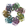
 Z (Sec.)
 Y (Row.)
 X (Col.)


 Sample components
 Processing
 Electron microscopy
 FIELD EMISSION GUN