Journal: J Mol Biol / Year: 2015 Title: Structure of Full-Length Human PDGFRβ Bound to Its Activating Ligand PDGF-B as Determined by Negative-Stain Electron Microscopy. Authors: Po-Han Chen / Vinzenz Unger / Xiaolin He / Abstract: Members of the receptor tyrosine kinases (RTKs) regulate important cellular functions such as cell growth and migration, which are key steps in angiogenesis, in organ morphogenesis and in the ...Members of the receptor tyrosine kinases (RTKs) regulate important cellular functions such as cell growth and migration, which are key steps in angiogenesis, in organ morphogenesis and in the unregulated states, cancer formation. One long-standing puzzle regarding RTKs centers on how the extracellular domain (ECD), which detects and binds to growth factors, is coupled with the intracellular domain kinase activation. While extensive structural works on the soluble portions of RTKs have provided critical insights into RTK structures and functions, lack of a full-length receptor structure has hindered a comprehensive overview of RTK activation. In this study, we successfully purified and determined a 27-Å-resolution structure of PDGFRβ [a full-length human platelet-derived growth factor receptor], in complex with its ligand PDGF-B. In the ligand-stimulated complex, two PDGFRβs assemble into a dimer via an extensive interface essentially running along the full-length of the receptor, suggesting that the membrane-proximal region, the transmembrane helix and the kinase domain of PDGFRβ are involved in dimerization. Major structural differences are seen between the full-length and soluble ECD structures, rationalizing previous experimental data on how membrane-proximal domains modulate receptor ligand-binding affinity and dimerization efficiency. Also, in contrast to the 2-fold symmetry of the ECD, the intracellular kinase domains adopt an asymmetric dimer arrangement, in agreement with prior observations for the closely related KIT receptor. In essence, the structure provides a first glimpse into how platelet-derived growth factor receptor ECD, upon ligand stimulation, is coupled to its intracellular domain kinase activation.
History
Deposition
Aug 12, 2015
-
Header (metadata) release
Sep 23, 2015
-
Map release
May 4, 2016
-
Update
May 4, 2016
-
Current status
May 4, 2016
Processing site: RCSB / Status: Released
-
Structure visualization
Movie
Surface view with section colored by density value
Protein or peptide: Platelet-derived growth factor receptor beta
Protein or peptide: Platelet-derived growth factor-BB
-
Supramolecule #1000: human PDGF-B bound to PDGFR-Beta
Supramolecule
Name: human PDGF-B bound to PDGFR-Beta / type: sample / ID: 1000 Details: The sample is cross-linked via the GraFix method after the gel filtration step. Oligomeric state: a PDGF-B dimer bound to two PDGFR-Beta / Number unique components: 2
Molecular weight
Experimental: 300 KDa / Theoretical: 300 KDa / Method: gel filtration and SDS-PAGE analysis
Name: Platelet-derived growth factor receptor beta / type: protein_or_peptide / ID: 1 / Name.synonym: PDGFR-Beta Details: N-terminal Flag tag is attached after a secretion signal derived from Gaussia luciferase. Number of copies: 2 / Oligomeric state: Dimer / Recombinant expression: Yes
Source (natural)
Organism: Homo sapiens (human) / synonym: Human / Location in cell: plasma membrane
Initial models are reconstructed by RCT. Visual inspection of similar classes were pooled, and one initial model was used as an input model for 3D classification into three classes using RELION.
Final reconstruction
Algorithm: OTHER / Resolution.type: BY AUTHOR / Resolution: 27.0 Å / Resolution method: OTHER / Software - Name: XMIPP, RELION / Number images used: 4234
+
About Yorodumi
-
News
-
Feb 9, 2022. New format data for meta-information of EMDB entries
New format data for meta-information of EMDB entries
Version 3 of the EMDB header file is now the official format.
The previous official version 1.9 will be removed from the archive.
In the structure databanks used in Yorodumi, some data are registered as the other names, "COVID-19 virus" and "2019-nCoV". Here are the details of the virus and the list of structure data.
Jan 31, 2019. EMDB accession codes are about to change! (news from PDBe EMDB page)
EMDB accession codes are about to change! (news from PDBe EMDB page)
The allocation of 4 digits for EMDB accession codes will soon come to an end. Whilst these codes will remain in use, new EMDB accession codes will include an additional digit and will expand incrementally as the available range of codes is exhausted. The current 4-digit format prefixed with “EMD-” (i.e. EMD-XXXX) will advance to a 5-digit format (i.e. EMD-XXXXX), and so on. It is currently estimated that the 4-digit codes will be depleted around Spring 2019, at which point the 5-digit format will come into force.
The EM Navigator/Yorodumi systems omit the EMD- prefix.
Related info.:Q: What is EMD? / ID/Accession-code notation in Yorodumi/EM Navigator
Yorodumi is a browser for structure data from EMDB, PDB, SASBDB, etc.
This page is also the successor to EM Navigator detail page, and also detail information page/front-end page for Omokage search.
The word "yorodu" (or yorozu) is an old Japanese word meaning "ten thousand". "mi" (miru) is to see.
Related info.:EMDB / PDB / SASBDB / Comparison of 3 databanks / Yorodumi Search / Aug 31, 2016. New EM Navigator & Yorodumi / Yorodumi Papers / Jmol/JSmol / Function and homology information / Changes in new EM Navigator and Yorodumi
 Movie
Movie Controller

 Yorodumi
 Open data
 Basic information
 Map data
 Sample
 Keywords
 Function and homology information
 Homo sapiens (human)
 Authors
 Citation
 Structure visualization
 Downloads & links
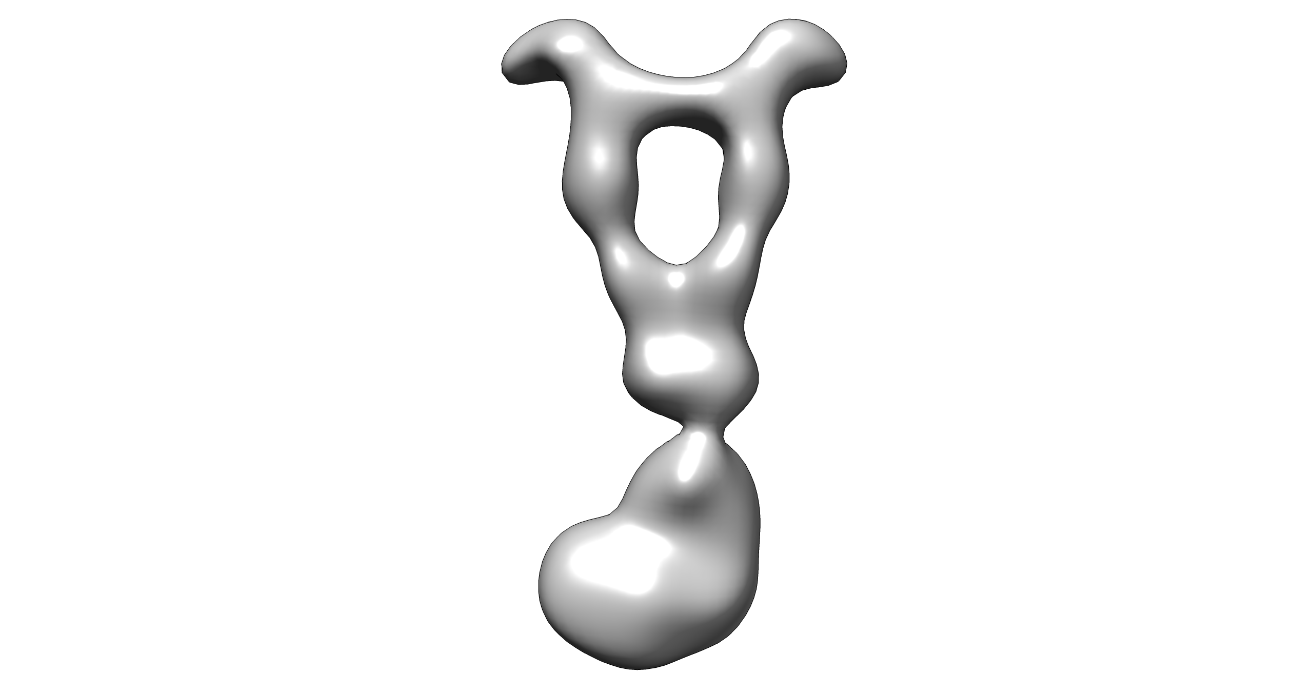 emd_6426.png
 http://ftp.pdbj.org/pub/emdb/structures/EMD-6426
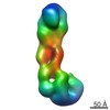


 Z (Sec.)
 Y (Row.)
 X (Col.)


 Sample components
 Processing
 Electron microscopy
 FIELD EMISSION GUN