 Movie
Movie Controller


+ Open data

- Basic information
| Entry |  | |||||||||
|---|---|---|---|---|---|---|---|---|---|---|
| Title | PsdAB dimer (peptidisc sample) | |||||||||
 Map data Map data | ||||||||||
 Sample Sample |
| |||||||||
 Keywords Keywords | ABC transpoter / TRANSPORT PROTEIN | |||||||||
| Function / homology | : / :  Function and homology information Function and homology information | |||||||||
| Biological species |  | |||||||||
| Method | single particle reconstruction / cryo EM / Resolution: 3.6 Å | |||||||||
 Authors Authors | He YT / Fan WJ / Shao K / Luo M | |||||||||
| Funding support |  Singapore, 1 items Singapore, 1 items
| |||||||||
 Citation Citation | Journal: Structure / Year: 2026 Title: Cryo-EM structure of the Nisin resistance pump PsdAB reveals an unusual ABC transporter architecture. Authors: Yutong He / Wenjie Fan / Jian Shi / Bee Koon Gan / Kai Shao / Fan Zhu / Xuechuan Hong / Min Luo /  Abstract: Bacteria have evolved diverse strategies to resist antimicrobial peptides, among them lipid II-targeting lantibiotics such as nisin. PsdAB, an ABC-type transporter regulated by the PsdRS two- ...Bacteria have evolved diverse strategies to resist antimicrobial peptides, among them lipid II-targeting lantibiotics such as nisin. PsdAB, an ABC-type transporter regulated by the PsdRS two-component system, contributes to nisin resistance, though its structural and mechanistic basis have remained unclear. Here, we report the cryo-EM structure of Bacillus subtilis PsdAB, revealing a dimeric assembly with an unusually large central cavity at the TMD interface. Cross-linking studies confirm the dimeric nature of PsdAB both in vitro and in cells. Functional assays demonstrate that dimer-disrupting mutations compromise nisin resistance, highlighting the importance of dimerization for activity. Compared to canonical ABC transporter types, PsdAB adopts an atypical architecture comprising four NBDs and two TMDs arranged around a central cavity, which may accommodate lipid II. We propose that PsdAB represents a previously unrecognized ABC transporter class. These findings offer new insights into transporter-mediated lantibiotic resistance and suggest a potential mechanism of lipid II shielding. | |||||||||
| History |
|
- Structure visualization
| Supplemental images |
|---|

- Downloads & links
-EMDB archive
| Map data | emd_64138.map.gz | 59.4 MB | EMDB map data format | |
|---|---|---|---|---|
| Header (meta data) | emd-64138-v30.xmlemd-64138.xml | 17.3 KB 17.3 KB | Display Display | EMDB header |
| FSC (resolution estimation) | emd_64138_fsc.xml | 8.5 KB | Display | FSC data file |
| Images |  emd_64138.png emd_64138.png | 19.1 KB | ||
| Filedesc metadata | emd-64138.cif.gz | 6 KB | ||
| Others | emd_64138_half_map_1.map.gzemd_64138_half_map_2.map.gz | 59.4 MB 59.4 MB | ||
| Archive directory |  http://ftp.pdbj.org/pub/emdb/structures/EMD-64138ftp://ftp.pdbj.org/pub/emdb/structures/EMD-64138 http://ftp.pdbj.org/pub/emdb/structures/EMD-64138ftp://ftp.pdbj.org/pub/emdb/structures/EMD-64138 | HTTPS FTP |
-Related structure data
| Related structure data | 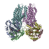 9uggMC  9wmiC M: atomic model generated by this map C: citing same article ( |
|---|---|
| Similar structure data |

-Links
| EMDB pages | EMDB (EBI/PDBe) / EMDataResource |
|---|
-Map
| File | Download / File: emd_64138.map.gz / Format: CCP4 / Size: 64 MB / Type: IMAGE STORED AS FLOATING POINT NUMBER (4 BYTES) | ||||||||||||||||||||||||||||||||||||
|---|---|---|---|---|---|---|---|---|---|---|---|---|---|---|---|---|---|---|---|---|---|---|---|---|---|---|---|---|---|---|---|---|---|---|---|---|---|
| Projections & slices | Image control
Images are generated by Spider. | ||||||||||||||||||||||||||||||||||||
| Voxel size | X=Y=Z: 1.06 Å | ||||||||||||||||||||||||||||||||||||
| Density |
| ||||||||||||||||||||||||||||||||||||
| Symmetry | Space group: 1 | ||||||||||||||||||||||||||||||||||||
| Details | EMDB XML:
|
 Z (Sec.)
 Y (Row.)
 X (Col.)


-Supplemental data
-Half map: #2
| File | emd_64138_half_map_1.map | ||||||||||||
|---|---|---|---|---|---|---|---|---|---|---|---|---|---|
| Projections & Slices |
| ||||||||||||
| Density Histograms |


-Half map: #1
| File | emd_64138_half_map_2.map | ||||||||||||
|---|---|---|---|---|---|---|---|---|---|---|---|---|---|
| Projections & Slices |
| ||||||||||||
| Density Histograms |


- Sample components
-Entire : Hexa complex of PsdA and PsdB.
| Entire | Name: Hexa complex of PsdA and PsdB. |
|---|---|
| Components |
|
-Supramolecule #1: Hexa complex of PsdA and PsdB.
| Supramolecule | Name: Hexa complex of PsdA and PsdB. / type: complex / ID: 1 / Parent: 0 / Macromolecule list: #1-#2 |
|---|---|
| Source (natural) | Organism: |
-Macromolecule #1: Lantibiotic ABC transporter ATP-binding protein PsdA
| Macromolecule | Name: Lantibiotic ABC transporter ATP-binding protein PsdA / type: protein_or_peptide / ID: 1 / Number of copies: 4 / Enantiomer: LEVO |
|---|---|
| Source (natural) | Organism: |
| Molecular weight | Theoretical: 29.073227 KDa |
| Recombinant expression | Organism: |
| Sequence | String: MNVLQTTNLS KTYYSNKGTI SYQALSAFDL SVSKGEFVGI MGPSGSGKTT LLNLLATIDK PTQGEMMING IQPKTLKDQE LALFRRREL GFVFQDFNLL DTLTIRENIL LPLALDKVKL REMEARLDEL ADTLQIKHIL DHRTYEVSGG QQQRAACARA I IHNPALIL ...String: MNVLQTTNLS KTYYSNKGTI SYQALSAFDL SVSKGEFVGI MGPSGSGKTT LLNLLATIDK PTQGEMMING IQPKTLKDQE LALFRRREL GFVFQDFNLL DTLTIRENIL LPLALDKVKL REMEARLDEL ADTLQIKHIL DHRTYEVSGG QQQRAACARA I IHNPALIL ADEPTGNLDS KSAKQVMNTL AQLNEEKEAT ILLVTHDATA ASFCKRIVFI KDGRFFSEIH RGTNRQVFYQ SI LDTLSVL GGDFHEFENY RP UniProtKB: UNIPROTKB: A0AA96UMI8 |
-Macromolecule #2: Lantibiotic ABC transporter permease PsdB
| Macromolecule | Name: Lantibiotic ABC transporter permease PsdB / type: protein_or_peptide / ID: 2 / Number of copies: 2 / Enantiomer: LEVO |
|---|---|
| Source (natural) | Organism: |
| Molecular weight | Theoretical: 73.589867 KDa |
| Recombinant expression | Organism: |
| Sequence | String: MNLRTIARKN ILGNLQRYVA YFLSCVFAVS VFFVFTSFIF HPDVNEDNIY GGSLVKTCLS AALVVIIVFC IFFITYSNSA FLQARKKEF GLLTLFGTSK QQLRKMIYYE QSLISLAAIA AGIGAGLLFS KLFFMIMTWM LSVKVPISFA IVPKAFVMTI A GFLILFQT ...String: MNLRTIARKN ILGNLQRYVA YFLSCVFAVS VFFVFTSFIF HPDVNEDNIY GGSLVKTCLS AALVVIIVFC IFFITYSNSA FLQARKKEF GLLTLFGTSK QQLRKMIYYE QSLISLAAIA AGIGAGLLFS KLFFMIMTWM LSVKVPISFA IVPKAFVMTI A GFLILFQT LLILSLGRIR KLEIIELIKS AKKPKSLPVY SKWLTVLSLL CLGSGYYLSA TANAIDMMFR VFPILILVLV GT YFFFTQS SVAFFRMLYR KKHSFYKGTN IIVRSNMIFR LKDHARMLFL TSVITAVILT ATGVIYMFYS DLQRQEEQSI PQS VSWVEK DASRFQVMKP ETAENTLKKA HAVIKYKVDA TGIPVTFQSD LPYGNKKMEA EALLISEKVY NQVAKEKGFP VIHL QENEA FINVSFQMMV KDTFGEGETA AFHMKSGKTL SYVMKKQQNK GILMSVDGVS RLLVVSEKSF DSLSQDVPLK EQMRM VGYE LEHWQETVDV SEKLENMVPK EHTSDFQTRA PSYQIVKQGV ALMLFIGLFV SVLFFIVQGS MLYLRMFTEI EDTRVQ VLA LKRIGVTDKE IHSILGKQIG FLFFIPFIAG TIHAGFAYAA LSNMLNSNLF LEAVIVIFIY FVFQALYYIV TRHIYKR AV LQRM UniProtKB: UNIPROTKB: A0AA96UNF6 |
-Macromolecule #3: ADENOSINE-5'-DIPHOSPHATE
| Macromolecule | Name: ADENOSINE-5'-DIPHOSPHATE / type: ligand / ID: 3 / Number of copies: 4 / Formula: ADP |
|---|---|
| Molecular weight | Theoretical: 427.201 Da |
| Chemical component information |  ChemComp-ADP: |
-Experimental details
-Structure determination
| Method | cryo EM |
|---|---|
 Processing Processing | single particle reconstruction |
| Aggregation state | particle |
-Sample preparation
| Buffer | pH: 7.5 |
|---|---|
| Vitrification | Cryogen name: ETHANE |
- Electron microscopy
| Microscope | TFS KRIOS |
|---|---|
| Image recording | Film or detector model: GATAN K3 (6k x 4k) / Average electron dose: 39.0 e/Å2 |
| Electron beam | Acceleration voltage: 300 kV / Electron source:  FIELD EMISSION GUN FIELD EMISSION GUN |
| Electron optics | Illumination mode: SPOT SCAN / Imaging mode: BRIGHT FIELD / Nominal defocus max: 2.5 µm / Nominal defocus min: 0.8 µm |
| Experimental equipment |  Model: Titan Krios / Image courtesy: FEI Company |

