 Movie
Movie Controller

[English] 日本語
 Yorodumi
- EMDB-63624: Cryo-EM structure of Dp42 depolymerase with C3 symmetry against K... -
+ Open data

- Basic information
| Entry |  | |||||||||
|---|---|---|---|---|---|---|---|---|---|---|
| Title | Cryo-EM structure of Dp42 depolymerase with C3 symmetry against KN1 serotype Klebsiella pneumoniae,(Dp42-C3) | |||||||||
 Map data Map data | Dp42 with C3 symmetry | |||||||||
 Sample Sample |
| |||||||||
 Keywords Keywords | Dp42 / depolymerase / cryo-EM / Klebsiella pneumoniae KN1 / capsule polysaccharide LYASE / LYASE | |||||||||
| Function / homology | symbiont entry into host cell via disruption of host cell glycocalyx / Bacteriophage T7 tail fibre protein / Phage T7 tail fibre protein, N-terminal domain / symbiont entry into host cell via disruption of host cell envelope / virus tail / adhesion receptor-mediated virion attachment to host cell / Probable tail spike protein Function and homology information Function and homology information | |||||||||
| Biological species |  Klebsiella phage vB_KpnP_IME321 (virus) Klebsiella phage vB_KpnP_IME321 (virus) | |||||||||
| Method | single particle reconstruction / cryo EM / Resolution: 2.56 Å | |||||||||
 Authors Authors | Xie Y / Huang T / Shi X / Tao X / Ma C | |||||||||
| Funding support |  China, 1 items China, 1 items
| |||||||||
 Citation Citation | Journal: To Be Published Title: Structures and the mechanism investigation of bacteriophage depolymerases for capsular polysaccharide degradation of Klebsiella pneumoniae KN1 serotype Authors: Xie Y | |||||||||
| History |
|
- Structure visualization
| Supplemental images |
|---|

- Downloads & links
-EMDB archive
| Map data | emd_63624.map.gz | 55.9 MB | EMDB map data format | |
|---|---|---|---|---|
| Header (meta data) | emd-63624-v30.xmlemd-63624.xml | 14.5 KB 14.5 KB | Display Display | EMDB header |
| FSC (resolution estimation) | emd_63624_fsc.xml | 8.9 KB | Display | FSC data file |
| Images |  emd_63624.png emd_63624.png | 33.5 KB | ||
| Filedesc metadata | emd-63624.cif.gz | 5.7 KB | ||
| Others | emd_63624_half_map_1.map.gzemd_63624_half_map_2.map.gz | 46.2 MB 46.2 MB | ||
| Archive directory |  https://data.pdbj.org/pub/emdb/structures/EMD-63624ftp://data.pdbj.org/pub/emdb/structures/EMD-63624 https://data.pdbj.org/pub/emdb/structures/EMD-63624ftp://data.pdbj.org/pub/emdb/structures/EMD-63624 | HTTPS FTP |
-Related structure data
| Related structure data |  9m4eMC  9m4cC  9m4dC M: atomic model generated by this map C: citing same article ( |
|---|---|
| Similar structure data |


-Links
| EMDB pages | EMDB (EBI/PDBe) / EMDataResource |
|---|
-Map
| File | Download / File: emd_63624.map.gz / Format: CCP4 / Size: 59.6 MB / Type: IMAGE STORED AS FLOATING POINT NUMBER (4 BYTES) | ||||||||||||||||||||||||||||||||||||
|---|---|---|---|---|---|---|---|---|---|---|---|---|---|---|---|---|---|---|---|---|---|---|---|---|---|---|---|---|---|---|---|---|---|---|---|---|---|
| Annotation | Dp42 with C3 symmetry | ||||||||||||||||||||||||||||||||||||
| Projections & slices | Image control
Images are generated by Spider. | ||||||||||||||||||||||||||||||||||||
| Voxel size | X=Y=Z: 0.851 Å | ||||||||||||||||||||||||||||||||||||
| Density |
| ||||||||||||||||||||||||||||||||||||
| Symmetry | Space group: 1 | ||||||||||||||||||||||||||||||||||||
| Details | EMDB XML:
|
 Z (Sec.)
 Y (Row.)
 X (Col.)


-Supplemental data
-Half map: EM half1 map of Dp42 with C3 symmetry
| File | emd_63624_half_map_1.map | ||||||||||||
|---|---|---|---|---|---|---|---|---|---|---|---|---|---|
| Annotation | EM half1 map of Dp42 with C3 symmetry | ||||||||||||
| Projections & Slices |
| ||||||||||||
| Density Histograms |


-Half map: EM half2 map of Dp42 with C3 symmetry
| File | emd_63624_half_map_2.map | ||||||||||||
|---|---|---|---|---|---|---|---|---|---|---|---|---|---|
| Annotation | EM half2 map of Dp42 with C3 symmetry | ||||||||||||
| Projections & Slices |
| ||||||||||||
| Density Histograms |


- Sample components
-Entire : Dp42 with C3 symmetry (Dp42-C3)
| Entire | Name: Dp42 with C3 symmetry (Dp42-C3) |
|---|---|
| Components |
|
-Supramolecule #1: Dp42 with C3 symmetry (Dp42-C3)
| Supramolecule | Name: Dp42 with C3 symmetry (Dp42-C3) / type: complex / ID: 1 / Parent: 0 / Macromolecule list: all |
|---|---|
| Source (natural) | Organism: Klebsiella phage vB_KpnP_IME321 (virus) |
-Macromolecule #1: Probable tail spike protein
| Macromolecule | Name: Probable tail spike protein / type: protein_or_peptide / ID: 1 / Number of copies: 3 / Enantiomer: LEVO |
|---|---|
| Source (natural) | Organism: Klebsiella phage vB_KpnP_IME321 (virus) |
| Molecular weight | Theoretical: 92.402719 KDa |
| Recombinant expression | Organism:   Escherichia coli BL21(DE3) (bacteria) Escherichia coli BL21(DE3) (bacteria) |
| Sequence | String: MGSSHHHHHH SSGLVPRGSH MNQDIKTIIQ YPVGDVEFDI PFDYLSRKFV RVYLVSPLNR RQLNNITEYR YVSRTRIKLL VETAGFNLI EIRRFTSASE RVVDFSDGSV LRAADLNVSQ LQSAHIAEEA RDAAMLTISP ADDGSLDASG KVIKNVGTPV Q SSDAATKG ...String: MGSSHHHHHH SSGLVPRGSH MNQDIKTIIQ YPVGDVEFDI PFDYLSRKFV RVYLVSPLNR RQLNNITEYR YVSRTRIKLL VETAGFNLI EIRRFTSASE RVVDFSDGSV LRAADLNVSQ LQSAHIAEEA RDAAMLTISP ADDGSLDASG KVIKNVGTPV Q SSDAATKG YVDTTVAPLA TTIEANFMRT LRTSGRSIRE LPGASEVAGM LLGFNGEGDP VPVVAGEGTA SDVMLKLAGT TG LSYIGGV GYVTPEMMTV DGKTLVRGLG QDHVRFIQKA IDEGHRRNVP VLLSGGGYEV YETLHDAPLP RDDGTAYPEW VAN GGDSNI RPEEQLYQKA HLRLYNNSVI LGAGSQITTI RSTWSRGTSA VDLTSPIMWY IEGPLGNRGT VSYVLKGIKT MGAY IGRYV VGISYRSIED DLEFSGCGIS GVKQGEEQTL HRKIVITAYA GDVTGGWWLQ RNNAYGTKYM PPYTDTDVWL MGWCD SSKY EYLSYTGYDY YGRDALVHDW ISEWFDTYIF KSANSRKVSE GGRLTNQSAN PYPLPTLKGI TGRARYITSR YSRQNA LNI IDTLKTLITI RAPGYMDNST QSCRIVNAMI EAVGLIRRTS GANAGNYFGI DVVDKWGADT GVWGLEGTGI LEQQLVV FL RPGVPCTNAV VATGAGQIFE SWTTTNAQRR LLALRDWNPA TQVQTYRYDF RTDYALMRPT RYYTDGPLWN YSKGTSTP T VAVNGSAIAV QKAVSNWYRL GDIMRCNIYV EINSITLQGN SELTVTTPSY NGSWEVAGQG IGKVYLSTLT GDVTLTPVI QQGSNVVRLR RGSSPEVYTF AAGTYNNVVL IIGIDYVPAS UniProtKB: Probable tail spike protein |
-Experimental details
-Structure determination
| Method | cryo EM |
|---|---|
 Processing Processing | single particle reconstruction |
| Aggregation state | particle |
-Sample preparation
| Buffer | pH: 8 |
|---|---|
| Vitrification | Cryogen name: ETHANE |
- Electron microscopy
| Microscope | FEI TALOS ARCTICA |
|---|---|
| Image recording | Film or detector model: GATAN K3 BIOQUANTUM (6k x 4k) / Average electron dose: 54.0 e/Å2 |
| Electron beam | Acceleration voltage: 300 kV / Electron source:  FIELD EMISSION GUN FIELD EMISSION GUN |
| Electron optics | Illumination mode: FLOOD BEAM / Imaging mode: BRIGHT FIELD / Nominal defocus max: 1.5 µm / Nominal defocus min: 1.1 µm |
| Experimental equipment | 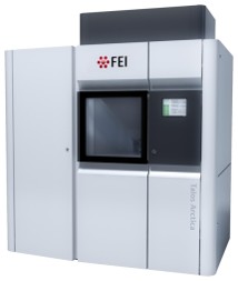 Model: Talos Arctica / Image courtesy: FEI Company |

