Movie
Movie Controller


+ Open data

- Basic information
| Entry |  | ||||||||||||||||||
|---|---|---|---|---|---|---|---|---|---|---|---|---|---|---|---|---|---|---|---|
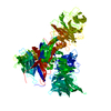
| Title | HBx R96E fused DDB1 4M mutant | ||||||||||||||||||
 Map data Map data | |||||||||||||||||||
 Sample Sample |
| ||||||||||||||||||
 Keywords Keywords | HBV / complex / VIRAL PROTEIN | ||||||||||||||||||
| Biological species |  Homo sapiens (human) Homo sapiens (human) | ||||||||||||||||||
| Method | single particle reconstruction / cryo EM / Resolution: 3.35 Å | ||||||||||||||||||
 Authors Authors | Tanaka H / Kita S / Sasaki M / Maenaka K / Machida S | ||||||||||||||||||
| Funding support |  Japan, 5 items Japan, 5 items
| ||||||||||||||||||
 Citation Citation | Journal: Proc Natl Acad Sci U S A / Year: 2025 Title: Structural basis of the hepatitis B virus X protein in complex with DDB1. Authors: Hiroki Tanaka / Joao Diogo Dias / Basile Jay / Shunsuke Kita / Mina Sasaki / Hiroyuki Takeda / Naoki Kishimoto / Shunsuke Sasaki / Shogo Misumi / Masashi Mizokami / Christine Neuveut / ...Authors: Hiroki Tanaka / Joao Diogo Dias / Basile Jay / Shunsuke Kita / Mina Sasaki / Hiroyuki Takeda / Naoki Kishimoto / Shunsuke Sasaki / Shogo Misumi / Masashi Mizokami / Christine Neuveut / Takashi Sumikama / Mikihiro Shibata / Katsumi Maenaka / Shinichi Machida /  Abstract: A cure for chronic hepatitis B requires eliminating or permanently silencing covalently closed circular DNA (cccDNA). A pivotal target of this approach is the hepatitis B virus (HBV) X protein (HBx), ...A cure for chronic hepatitis B requires eliminating or permanently silencing covalently closed circular DNA (cccDNA). A pivotal target of this approach is the hepatitis B virus (HBV) X protein (HBx), which is a key factor that promotes transcription from cccDNA. However, the HBx structure remains unsolved. Here, we present the cryoelectron microscopy structure of HBx in complex with DDB1, which is an essential complex for cccDNA transcription. In this structure, hydrophobic interactions within HBx were identified, and mutational analysis highlighted their importance in the HBV life cycle. Our biochemical analysis revealed that the HBx-DDB1 complex directly interacts simultaneously with NSE3, which is a component of the SMC5/6 complex, and Spindlin1. Additionally, HBx-DDB1 complex dynamics were explored via high-speed atomic force microscopy. These findings provide comprehensive insights into the structure and function of HBx in HBV replication. | ||||||||||||||||||
| History |
|
- Structure visualization
| Supplemental images |
|---|

- Downloads & links
-EMDB archive
| Map data | emd_63366.map.gz | 8.8 MB |  EMDB map data format EMDB map data format | |
|---|---|---|---|---|
| Header (meta data) | emd-63366-v30.xmlemd-63366.xml | 15.9 KB 15.9 KB | Display Display | EMDB header |
| Images |  emd_63366.png emd_63366.png | 70.8 KB | ||
| Filedesc metadata | emd-63366.cif.gz | 5.7 KB | ||
| Others | emd_63366_half_map_1.map.gzemd_63366_half_map_2.map.gz | 98.3 MB 98.4 MB | ||
| Archive directory |  http://ftp.pdbj.org/pub/emdb/structures/EMD-63366ftp://ftp.pdbj.org/pub/emdb/structures/EMD-63366 http://ftp.pdbj.org/pub/emdb/structures/EMD-63366ftp://ftp.pdbj.org/pub/emdb/structures/EMD-63366 | HTTPS FTP |
-Related structure data


-Links
| EMDB pages | EMDB (EBI/PDBe) / EMDataResource |
|---|
-Map
| File | Download / File: emd_63366.map.gz / Format: CCP4 / Size: 125 MB / Type: IMAGE STORED AS FLOATING POINT NUMBER (4 BYTES) | ||||||||||||||||||||||||||||||||||||
|---|---|---|---|---|---|---|---|---|---|---|---|---|---|---|---|---|---|---|---|---|---|---|---|---|---|---|---|---|---|---|---|---|---|---|---|---|---|
| Projections & slices | Image control
Images are generated by Spider. | ||||||||||||||||||||||||||||||||||||
| Voxel size | X=Y=Z: 0.67 Å | ||||||||||||||||||||||||||||||||||||
| Density |
| ||||||||||||||||||||||||||||||||||||
| Symmetry | Space group: 1 | ||||||||||||||||||||||||||||||||||||
| Details | EMDB XML:
|
 Z (Sec.)
 Y (Row.)
 X (Col.)


-Supplemental data
-Half map: #1
| File | emd_63366_half_map_1.map | ||||||||||||
|---|---|---|---|---|---|---|---|---|---|---|---|---|---|
| Projections & Slices |
| ||||||||||||
| Density Histograms |


-Half map: #2
| File | emd_63366_half_map_2.map | ||||||||||||
|---|---|---|---|---|---|---|---|---|---|---|---|---|---|
| Projections & Slices |
| ||||||||||||
| Density Histograms |


- Sample components
-Entire : the complex of HBx and DDB1
| Entire | Name: the complex of HBx and DDB1 |
|---|---|
| Components |
|
-Supramolecule #1: the complex of HBx and DDB1
| Supramolecule | Name: the complex of HBx and DDB1 / type: complex / ID: 1 / Parent: 0 / Macromolecule list: all |
|---|---|
| Source (natural) | Organism: Homo sapiens (human) |
-Macromolecule #1: Protein X, DNA damage-binding protein 1
| Macromolecule | Name: Protein X, DNA damage-binding protein 1 / type: protein_or_peptide / ID: 1 / Enantiomer: LEVO |
|---|---|
| Source (natural) | Organism: Homo sapiens (human) |
| Recombinant expression | Organism: Homo sapiens (human) |
| Sequence | String: MASAWSHPQF EKGSGSGMAA RLYCQLDPSR DVLCLRPVGA ESRGRPLSGP LGTLSSPSPS AVPADHGAHL SLRGLPVCAF SSAGPCALRF TSARCMETTV NAHQILPKVL HKETLGLPAM STTDLEAYFK DCVFKDWEEL GEEIRLKVFV LGGCRHKLVC APAPCNFFTS ...String: MASAWSHPQF EKGSGSGMAA RLYCQLDPSR DVLCLRPVGA ESRGRPLSGP LGTLSSPSPS AVPADHGAHL SLRGLPVCAF SSAGPCALRF TSARCMETTV NAHQILPKVL HKETLGLPAM STTDLEAYFK DCVFKDWEEL GEEIRLKVFV LGGCRHKLVC APAPCNFFTS ASGSGSGSGS GSGMSYNYVV TAQKPTAVNG CVTGHFTSAE DLNLLIAKNT RLEIYVVTAE GLRPVKEVGM YGKIAVMELF RPKGESKDLL FILTAKYNAC ILEYKQSGES IDIITRAHGN VQDRIGRPSE TGIIGIIDPE CRMIGLRLYD GLFKVIPLDR DNKELKAFNI RLEELHVIDV KFLYGCQAPT ICFVYQDPQG RHVKTYEVSL REKEFNKGPW KQENVEAEAS MVIAVPEPFG GAIIIGQESI TYHNGDKYLA IAPPIIKQST IVCHNRVDPN GSRYLLGDME GRLFMLLLEK EEQMDGTVTL KDLRVELLGE TSIAECLTYL DNGVVFVGSR LGDSQLVKLN VDSNEQGSYV VAMETFTNLG PIVDMCVVDL ERQGQGQLVT CSGAFKEGSL RIIRNGIGIH EHDSIDLPGI KGLWPLRSDP NRETDDTLVL SFVGQTRVLM LNGEEVEETE LMGFVDDQQT FFCGNVAHQQ LIQITSASVR LVSQEPKALV SEWKEPQAKN ISVASCNSSQ VVVAVGRALY YLQIHPQELR QISHTEMEHE VACLDITPLG DSNGLSPLCA IGLKTDISAR ILKLPSFELL HKEMLGGEID PESILMTTFE SSHYLLCALG DGALFYFGLN IETGLLSDRK KVTLGTQPTV LRTFRSLSTT NVFACSDRPT VIYSSNHKLV FSNVNLKEVN YMCPLNSDGY PDSLALANNS TLTIGTIDEI QKLHIRTVPL YESPRKICYQ EVSQCFGVLS SRIEVQDTSG GTTALRPSAS TQALSSSVSS SKLFSSSTAP HETSFGEEVE VHNLLIIDQH TFEVLHAHQF LQNEYALSLV SCKLGKDPNT YFIVGTAMVY PEEAEPKQGR IVVFQYSDGK LQTVAEKEVK GAVYSMVEFN GKLLASINST VRLYEWTTEK ELRTECNHYN NIMALYLKTK GDFILVGDLM RSVLLLAYKP MEGNFEEIAR DFNPNWMSAV EILDDDNFLG AENAFNLFVC QKDSAATTDE ERQHLQEVGL FHLGEFVNVF CHGSLVMQNL GETSTPTQGS VLFGTVNGMI GLVTSLSESW YNLLLDMQNR LNKVIKSVGK IEHSFWRSFH TERKTEPATG FIDGDLIESF LDISRPKMQE VVANLQYDDG SGMKREATAD DLIKVVEELT RIH |
-Experimental details
-Structure determination
| Method | cryo EM |
|---|---|
 Processing Processing | single particle reconstruction |
| Aggregation state | particle |
-Sample preparation
| Buffer | pH: 7.5 |
|---|---|
| Vitrification | Cryogen name: ETHANE / Instrument: LEICA EM GP |
- Electron microscopy
| Microscope | TFS KRIOS |
|---|---|
| Specialist optics | Energy filter - Name: GIF Bioquantum / Energy filter - Slit width: 20 eV |
| Image recording | Film or detector model: GATAN K3 BIOQUANTUM (6k x 4k) / Average electron dose: 49.6 e/Å2 |
| Electron beam | Acceleration voltage: 300 kV / Electron source:  FIELD EMISSION GUN FIELD EMISSION GUN |
| Electron optics | Illumination mode: FLOOD BEAM / Imaging mode: BRIGHT FIELD / Nominal defocus max: 2.4 µm / Nominal defocus min: 0.8 µm |
| Experimental equipment |  Model: Titan Krios / Image courtesy: FEI Company |