 Movie
Movie Controller

[English] 日本語
 Yorodumi
- EMDB-6328: 3D reconstruction from 188 out of 450 fibrils of Abeta(1-40), sel... -
+ Open data

- Basic information
| Entry | Database: EMDB / ID: EMD-6328 | |||||||||
|---|---|---|---|---|---|---|---|---|---|---|
| Title | 3D reconstruction from 188 out of 450 fibrils of Abeta(1-40), selected by mean crossover-to-crossover distance (130 to 150 nm) and curvature (straightest 188 filaments were selected) | |||||||||
 Map data Map data | 3D reconstruction of Abeta(1-40) fibril before any filtering, symmetrization, or masking. Figure 5D of the primary citation was generated from this map by application of helical symmetry and a B factor of -600A^2, with low-pass filtering to 7.5A. | |||||||||
 Sample Sample |
| |||||||||
| Biological species |  Homo sapiens (human) Homo sapiens (human) | |||||||||
| Method | helical reconstruction / cryo EM / Resolution: 7.1 Å | |||||||||
 Authors Authors | Rohou A / Grigorieff N | |||||||||
 Citation Citation | Journal: Proc Natl Acad Sci U S A / Year: 2008 Title: Paired beta-sheet structure of an Abeta(1-40) amyloid fibril revealed by electron microscopy. Authors: Carsten Sachse / Marcus Fändrich / Nikolaus Grigorieff /  Abstract: Alzheimer's disease is a neurodegenerative disorder that is characterized by the cerebral deposition of amyloid fibrils formed by Abeta peptide. Despite their prevalence in Alzheimer's and other ...Alzheimer's disease is a neurodegenerative disorder that is characterized by the cerebral deposition of amyloid fibrils formed by Abeta peptide. Despite their prevalence in Alzheimer's and other neurodegenerative diseases, important details of the structure of amyloid fibrils remain unknown. Here, we present a three-dimensional structure of a mature amyloid fibril formed by Abeta(1-40) peptide, determined by electron cryomicroscopy at approximately 8-A resolution. The fibril consists of two protofilaments, each containing approximately 5-nm-long regions of beta-sheet structure. A local twofold symmetry within each region suggests that pairs of beta-sheets are formed from equivalent parts of two Abeta(1-40) peptides contained in each protofilament. The pairing occurs via tightly packed interfaces, reminiscent of recently reported steric zipper structures. However, unlike these previous structures, the beta-sheet pairing is observed within an amyloid fibril and includes significantly longer amino acid sequences. | |||||||||
| History |
|
- Structure visualization
| Movie |
 Movie viewer Movie viewer |
|---|---|
| Structure viewer | EM map: SurfViewMolmilJmol/JSmol |
| Supplemental images |

- Downloads & links
-EMDB archive
| Map data | emd_6328.map.gz | 365.2 MB | EMDB map data format | |
|---|---|---|---|---|
| Header (meta data) | emd-6328-v30.xmlemd-6328.xml | 10.1 KB 10.1 KB | Display Display | EMDB header |
| FSC (resolution estimation) | emd_6328_fsc.xml | 17.9 KB | Display | FSC data file |
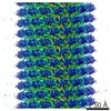
| Images | 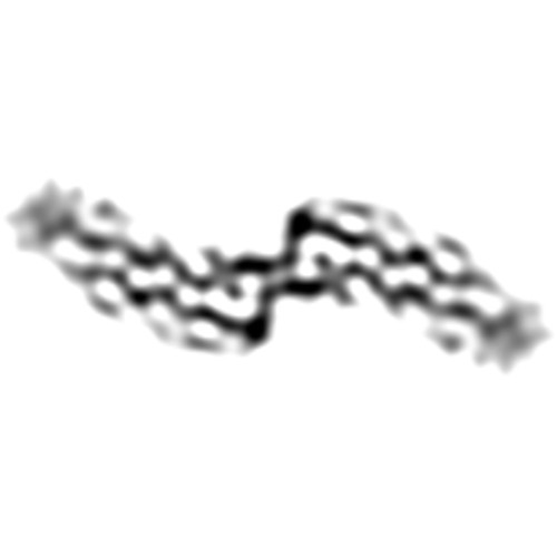 emd_6328.jpg emd_6328.jpg | 11.3 KB | ||
| Archive directory |  http://ftp.pdbj.org/pub/emdb/structures/EMD-6328ftp://ftp.pdbj.org/pub/emdb/structures/EMD-6328 http://ftp.pdbj.org/pub/emdb/structures/EMD-6328ftp://ftp.pdbj.org/pub/emdb/structures/EMD-6328 | HTTPS FTP |
-Related structure data

-Links
| EMDB pages | EMDB (EBI/PDBe) / EMDataResource |
|---|---|
| Related items in Molecule of the Month |

-Map
| File | Download / File: emd_6328.map.gz / Format: CCP4 / Size: 494.2 MB / Type: IMAGE STORED AS FLOATING POINT NUMBER (4 BYTES) | ||||||||||||||||||||||||||||||||||||||||||||||||||||||||||||
|---|---|---|---|---|---|---|---|---|---|---|---|---|---|---|---|---|---|---|---|---|---|---|---|---|---|---|---|---|---|---|---|---|---|---|---|---|---|---|---|---|---|---|---|---|---|---|---|---|---|---|---|---|---|---|---|---|---|---|---|---|---|
| Annotation | 3D reconstruction of Abeta(1-40) fibril before any filtering, symmetrization, or masking. Figure 5D of the primary citation was generated from this map by application of helical symmetry and a B factor of -600A^2, with low-pass filtering to 7.5A. | ||||||||||||||||||||||||||||||||||||||||||||||||||||||||||||
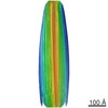
| Projections & slices | Image control
Images are generated by Spider. | ||||||||||||||||||||||||||||||||||||||||||||||||||||||||||||
| Voxel size | X=Y=Z: 1.2 Å | ||||||||||||||||||||||||||||||||||||||||||||||||||||||||||||
| Density |
| ||||||||||||||||||||||||||||||||||||||||||||||||||||||||||||
| Symmetry | Space group: 1 | ||||||||||||||||||||||||||||||||||||||||||||||||||||||||||||
| Details | EMDB XML:
CCP4 map header:
| ||||||||||||||||||||||||||||||||||||||||||||||||||||||||||||
 Z (Sec.)
 Y (Row.)
 X (Col.)


-Supplemental data
- Sample components
-Entire : human Abeta(1-40)
| Entire | Name: human Abeta(1-40) |
|---|---|
| Components |
|
-Supramolecule #1000: human Abeta(1-40)
| Supramolecule | Name: human Abeta(1-40) / type: sample / ID: 1000 / Oligomeric state: Helical assembly / Number unique components: 1 |
|---|
-Macromolecule #1: Amyloid beta peptide (1-40)
| Macromolecule | Name: Amyloid beta peptide (1-40) / type: protein_or_peptide / ID: 1 / Name.synonym: Abeta(1-40) / Recombinant expression: No / Database: NCBI |
|---|---|
| Source (natural) | Organism: Homo sapiens (human) / synonym: human |
-Experimental details
-Structure determination
| Method | cryo EM |
|---|---|
 Processing Processing | helical reconstruction |
| Aggregation state | filament |
-Sample preparation
| Concentration | 1 mg/mL |
|---|---|
| Buffer | pH: 7.8 / Details: 50 mM borate |
| Grid | Details: Quantifoil R1.2/1.3 Cu 400 mesh grids |
| Vitrification | Cryogen name: ETHANE / Instrument: HOMEMADE PLUNGER / Details: Vitrification carried out in cold room at 277 K. / Method: Blot for 7 seconds before plunging |
- Electron microscopy
| Microscope | FEI TECNAI F30 |
|---|---|
| Date | Feb 9, 2006 |
| Image recording | Category: FILM / Film or detector model: KODAK SO-163 FILM / Digitization - Scanner: ZEISS SCAI / Digitization - Sampling interval: 7.0 µm / Number real images: 63 / Average electron dose: 35 e/Å2 |
| Electron beam | Acceleration voltage: 300 kV / Electron source:  FIELD EMISSION GUN FIELD EMISSION GUN |
| Electron optics | Illumination mode: FLOOD BEAM / Imaging mode: BRIGHT FIELD / Cs: 2.0 mm / Nominal defocus max: 3.629 µm / Nominal defocus min: 2.027 µm / Nominal magnification: 59000 |
| Sample stage | Specimen holder model: GATAN LIQUID NITROGEN |
| Experimental equipment |  Model: Tecnai F30 / Image courtesy: FEI Company |
-Image processing
| Details | Filaments were processed using Frealix, including restraints on all refinement parameters. Data up to 20A were used in early stages of refinement. This limit was gradually increased to 9A. |
|---|---|
| Final reconstruction | Applied symmetry - Helical parameters - Δz: 4.8 Å Applied symmetry - Helical parameters - Δ&Phi: 0.734 ° Applied symmetry - Helical parameters - Axial symmetry: C2 (2 fold cyclic) Algorithm: OTHER / Resolution.type: BY AUTHOR / Resolution: 7.1 Å / Resolution method: OTHER / Software - Name: Frealix Details: Data up to 20A were used in early stages of the refinement. This limit was gradually increased to 9A. |
| CTF correction | Details: Defocus estimated for each helical subunit |
| FSC plot (resolution estimation) | 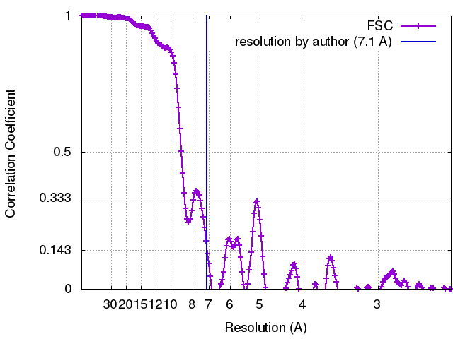 |
