Journal: mBio / Year: 2025 Title: Structural insights into distinct filamentation states reveal a regulatory mechanism for bacterial STING activation. Authors: Yuchao Yang / Yueyue Liu / Xue Ma / Xuan Zhao / Jian Cao / Yu Liu / Shanqin Li / Jing Wu / Yuanzhu Gao / Lianwan Chen / Changxin Wu / Guijun Shang / Sheng Liu / Defen Lu / Abstract: The cyclic oligonucleotide-based antiphage signaling system (CBASS) is a bacterial immune mechanism that was evolutionarily linked to the eukaryotic cGAS-STING pathway, which protects against phage ...The cyclic oligonucleotide-based antiphage signaling system (CBASS) is a bacterial immune mechanism that was evolutionarily linked to the eukaryotic cGAS-STING pathway, which protects against phage infection through abortive cell death. CBASS operons encode cyclic dinucleotide synthases (CD-NTases) and effector proteins (Caps), such as bacterial STING, which senses cyclic dinucleotides like 3'3'-c-di-GMP to trigger defense. Although bacterial STING oligomerizes into filaments upon ligand binding, the functional roles of distinct filament states remain unclear. Here, we resolve cryo-EM structures of TIR-STING (STING) bound to 3'3'-c-di-GMP, revealing two oligomeric states: spiral-shaped single filaments and fiber bundles composed of straight protofibrils. In spiral filaments, the STING domain sequesters the TIR domain's BB loop within a hydrophobic core, suppressing NADase activity. This inactive conformation is stabilized by interactions between the CBDα4 helix and the TIR domain, as well as a calcium-binding site. Conversely, fiber bundle formation-driven by inter-protofibril TIR domain interactions-disrupts these autoinhibitory contacts, liberating the BB loop to enable head-to-tail assembly of adjacent TIR domains into a composite NADase-active site. Calcium ions promote spiral filament assembly while inhibiting fiber bundles, revealing a dual regulatory role in tuning STING activation. Strikingly, this mechanism diverges from single-filament systems like STING, underscoring evolutionary diversity in STING signaling. Our findings establish distinct filament architectures as structural checkpoints governing bacterial STING activation, providing mechanistic insights into how conformational plasticity and environmental cues like calcium regulate abortive infection. These results highlight parallels between prokaryotic and eukaryotic immune strategies, emphasizing conserved principles in pathogen defense across domains of life.IMPORTANCEBacteria employ a sophisticated immune system, CBASS, evolutionarily related to human antiviral pathways, to defend against viral (phage) attacks. This study reveals how the bacterial protein STING acts as a molecular switch, transitioning between an inactive spiral structure stabilized by calcium ions and an active fiber bundle. When calcium levels drop, STING reorganizes into fiber bundles, activating its ability to degrade essential cellular molecules. This self-destructive mechanism halts phage replication by sacrificing the infected cell, protecting the bacterial population. The findings demonstrate how structural rearrangements govern life-or-death immune decisions, mirroring principles in human STING signaling. By uncovering calcium's role in regulating this process, the work deepens our understanding of microbial immunity and highlights shared strategies across domains of life. These insights could inspire novel antimicrobial therapies or bioengineered systems to combat infections, bridging fundamental science with practical applications in health and biotechnology.
In the structure databanks used in Yorodumi, some data are registered as the other names, "COVID-19 virus" and "2019-nCoV". Here are the details of the virus and the list of structure data.
Jan 31, 2019. EMDB accession codes are about to change! (news from PDBe EMDB page)
EMDB accession codes are about to change! (news from PDBe EMDB page)
The allocation of 4 digits for EMDB accession codes will soon come to an end. Whilst these codes will remain in use, new EMDB accession codes will include an additional digit and will expand incrementally as the available range of codes is exhausted. The current 4-digit format prefixed with “EMD-” (i.e. EMD-XXXX) will advance to a 5-digit format (i.e. EMD-XXXXX), and so on. It is currently estimated that the 4-digit codes will be depleted around Spring 2019, at which point the 5-digit format will come into force.
The EM Navigator/Yorodumi systems omit the EMD- prefix.
Related info.:Q: What is EMD? / ID/Accession-code notation in Yorodumi/EM Navigator
Yorodumi is a browser for structure data from EMDB, PDB, SASBDB, etc.
This page is also the successor to EM Navigator detail page, and also detail information page/front-end page for Omokage search.
The word "yorodu" (or yorozu) is an old Japanese word meaning "ten thousand". "mi" (miru) is to see.
Related info.:EMDB / PDB / SASBDB / Comparison of 3 databanks / Yorodumi Search / Aug 31, 2016. New EM Navigator & Yorodumi / Yorodumi Papers / Jmol/JSmol / Function and homology information / Changes in new EM Navigator and Yorodumi
 Movie
Movie Controller


 Open data
 Basic information
 Map data
 Sample
 Keywords
 Function and homology information
 Epilithonimonas lactis (bacteria)
 Authors
 China, 1 items
  Citation
 Structure visualization
 Downloads & links
 emd_63221.png
 http://ftp.pdbj.org/pub/emdb/structures/EMD-63221


 Z (Sec.)
 Y (Row.)
 X (Col.)


 Sample components
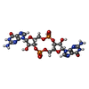

 Processing
 Electron microscopy
 FIELD EMISSION GUN
