+ Open data
- Basic information
| Entry |  | |||||||||
|---|---|---|---|---|---|---|---|---|---|---|
| Title | Overall structure of HKU5 S protein in closed conformation | |||||||||
 Map data Map data | ||||||||||
 Sample Sample |
| |||||||||
 Keywords Keywords | HKU5 / VIRAL PROTEIN | |||||||||
| Function / homology |  Function and homology information Function and homology informationhost cell endoplasmic reticulum-Golgi intermediate compartment membrane / receptor-mediated virion attachment to host cell / endocytosis involved in viral entry into host cell / fusion of virus membrane with host plasma membrane / fusion of virus membrane with host endosome membrane / viral envelope / host cell plasma membrane / virion membrane / membrane Similarity search - Function | |||||||||
| Biological species |  Bat coronavirus HKU5 Bat coronavirus HKU5 | |||||||||
| Method | single particle reconstruction / cryo EM / Resolution: 2.39 Å | |||||||||
 Authors Authors | Zhang YY / Xia LY / Zhou Q | |||||||||
| Funding support |  China, 1 items China, 1 items
| |||||||||
 Citation Citation |  Journal: To Be Published Journal: To Be PublishedTitle: Molecular Insights into Species-Specific ACE2 Recognition of Coronavirus HKU5 Authors: Zhang YY / Xia LY / Zhou Q | |||||||||
| History |
|
- Structure visualization
| Supplemental images |
|---|
- Downloads & links
-EMDB archive
| Map data |  emd_62522.map.gz emd_62522.map.gz | 118 MB |  EMDB map data format EMDB map data format | |
|---|---|---|---|---|
| Header (meta data) |  emd-62522-v30.xml emd-62522-v30.xml emd-62522.xml emd-62522.xml | 16.8 KB 16.8 KB | Display Display |  EMDB header EMDB header |
| Images |  emd_62522.png emd_62522.png | 71.2 KB | ||
| Filedesc metadata |  emd-62522.cif.gz emd-62522.cif.gz | 6.4 KB | ||
| Others |  emd_62522_half_map_1.map.gz emd_62522_half_map_1.map.gz emd_62522_half_map_2.map.gz emd_62522_half_map_2.map.gz | 116 MB 116 MB | ||
| Archive directory |  http://ftp.pdbj.org/pub/emdb/structures/EMD-62522 http://ftp.pdbj.org/pub/emdb/structures/EMD-62522 ftp://ftp.pdbj.org/pub/emdb/structures/EMD-62522 ftp://ftp.pdbj.org/pub/emdb/structures/EMD-62522 | HTTPS FTP |
-Validation report
| Summary document |  emd_62522_validation.pdf.gz emd_62522_validation.pdf.gz | 881.6 KB | Display |  EMDB validaton report EMDB validaton report |
|---|---|---|---|---|
| Full document |  emd_62522_full_validation.pdf.gz emd_62522_full_validation.pdf.gz | 881.2 KB | Display | |
| Data in XML |  emd_62522_validation.xml.gz emd_62522_validation.xml.gz | 14 KB | Display | |
| Data in CIF |  emd_62522_validation.cif.gz emd_62522_validation.cif.gz | 16.7 KB | Display | |
| Arichive directory |  https://ftp.pdbj.org/pub/emdb/validation_reports/EMD-62522 https://ftp.pdbj.org/pub/emdb/validation_reports/EMD-62522 ftp://ftp.pdbj.org/pub/emdb/validation_reports/EMD-62522 ftp://ftp.pdbj.org/pub/emdb/validation_reports/EMD-62522 | HTTPS FTP |
-Related structure data
| Related structure data |  9kr8MC  9kr9C  9kraC  9krbC M: atomic model generated by this map C: citing same article ( |
|---|---|
| Similar structure data | Similarity search - Function & homology  F&H Search F&H Search |
- Links
Links
| EMDB pages |  EMDB (EBI/PDBe) / EMDB (EBI/PDBe) /  EMDataResource EMDataResource |
|---|
- Map
Map
| File |  Download / File: emd_62522.map.gz / Format: CCP4 / Size: 125 MB / Type: IMAGE STORED AS FLOATING POINT NUMBER (4 BYTES) Download / File: emd_62522.map.gz / Format: CCP4 / Size: 125 MB / Type: IMAGE STORED AS FLOATING POINT NUMBER (4 BYTES) | ||||||||||||||||||||||||||||||||||||
|---|---|---|---|---|---|---|---|---|---|---|---|---|---|---|---|---|---|---|---|---|---|---|---|---|---|---|---|---|---|---|---|---|---|---|---|---|---|
| Projections & slices | Image control
Images are generated by Spider. | ||||||||||||||||||||||||||||||||||||
| Voxel size | X=Y=Z: 1.087 Å | ||||||||||||||||||||||||||||||||||||
| Density |
| ||||||||||||||||||||||||||||||||||||
| Symmetry | Space group: 1 | ||||||||||||||||||||||||||||||||||||
| Details | EMDB XML:
|
-Supplemental data
-Half map: #2
| File | emd_62522_half_map_1.map | ||||||||||||
|---|---|---|---|---|---|---|---|---|---|---|---|---|---|
| Projections & Slices |
| ||||||||||||
| Density Histograms |
-Half map: #1
| File | emd_62522_half_map_2.map | ||||||||||||
|---|---|---|---|---|---|---|---|---|---|---|---|---|---|
| Projections & Slices |
| ||||||||||||
| Density Histograms |
- Sample components
-Entire : Overall structure of HKU5 S protein in closed conformation
| Entire | Name: Overall structure of HKU5 S protein in closed conformation |
|---|---|
| Components |
|
-Supramolecule #1: Overall structure of HKU5 S protein in closed conformation
| Supramolecule | Name: Overall structure of HKU5 S protein in closed conformation type: complex / ID: 1 / Parent: 0 / Macromolecule list: #1 |
|---|---|
| Source (natural) | Organism:  Bat coronavirus HKU5 Bat coronavirus HKU5 |
-Macromolecule #1: Spike glycoprotein
| Macromolecule | Name: Spike glycoprotein / type: protein_or_peptide / ID: 1 / Number of copies: 3 / Enantiomer: LEVO |
|---|---|
| Source (natural) | Organism:  Bat coronavirus HKU5 Bat coronavirus HKU5 |
| Molecular weight | Theoretical: 149.814 KDa |
| Recombinant expression | Organism:  Homo sapiens (human) Homo sapiens (human) |
| Sequence | String: MIRSVLVLMC SLTFIGNLTR GQSVDMGHNG TGSCLDSQVQ PDYFESVHTT WPMPIDTSKA EGVIYPNGKS YSNITLTYTG LYPKANDLG KQYLFSDGHS APGRLNNLFV SNYSSQVESF DDGFVVRIGA AANKTGTTVI SQSTFKPIKK IYPAFLLGHS V GNYTPSNR ...String: MIRSVLVLMC SLTFIGNLTR GQSVDMGHNG TGSCLDSQVQ PDYFESVHTT WPMPIDTSKA EGVIYPNGKS YSNITLTYTG LYPKANDLG KQYLFSDGHS APGRLNNLFV SNYSSQVESF DDGFVVRIGA AANKTGTTVI SQSTFKPIKK IYPAFLLGHS V GNYTPSNR TGRYLNHTLV ILPDGCGTIL HAFYCVLHPR TQQNCAGETN FKSLSLWDTP ASDCVSGSYN QEATLGAFKV YF DLINCTF RYNYTITEDE NAEWFGITQD TQGVHLYSSR KENVFRNNMF HFATLPVYQK ILYYTVIPRS IRSPFNDRKA WAA FYIYKL HPLTYLLNFD VEGYITKAVD CGYDDLAQLQ CSYESFEVET GVYSVSSFEA SPRGEFIEQA TTQECDFTPM LTGT PPPIY NFKRLVFTNC NYNLTKLLSL FQVSEFSCHQ VSPSSLATGC YSSLTVDYFA YSTDMSSYLQ PGSAGAIVQF NYKQD FSNP TCRVLATVPQ NLTTITKPSN YAYLTECYKT SAYGKNYLYN APGAYTPCLS LASRGFSTKY QSHSDGELTT TGYIYP VTG NLQMAFIISV QYGTDTNSVC PMQALRNDTS IEDKLDVCVE YSLHGITGRG VFHNCTSVGL RNQRFVYDTF DNLVGYH SD NGNYYCVRPC VSVPVSVIYD KASNSHATLF GSVACSHVTT MMSQFSRMTK TNLLARTTPG PLQTTVGCAM GFINSSMV V DECQLPLGQS LCAIPPTTSS RVRRATSGAS DVFQIATLNF TSPLTLAPIN STGFVVAVPT NFTFGVTQEF IETTIQKIT VDCKQYVCNG FKKCEDLLKE YGQFCSKINQ ALHGANLRQD ESIANLFSSI KTQNTQPLQA GLNGDFNLTM LQIPQVTTGE RKYRSTIED LLFNKVTIAD PGYMQGYDEC MQQGPQSARD LICAQYVAGY KVLPPLYDPY MEAAYTSSLL GSIAGASWTA G LSSFAAIP FAQSIFYRLN GVGITQQVLS ENQKIIANKF NQALGAMQTG FTTTNLAFNK VQDAVNANAM ALSKLAAELS NT FGAISSS ISDILARLDT VEQEAQIDRL INGRLTSLNA FVAQQLVRTE AAARSAQLAQ DKVNECVKSQ SKRNGFCGTG THI VSFAIN APNGLYFFHV GYQPTSHVNA TAAYGLCNTE NPQKCIAPID GYFVLNQTTS TVADSDQQWY YTGSSFFHPE PITE ANSKY VSMDVKFENL TNRLPPPLLS NSTDLDFKEE LEEFFKNVSS QGPNFQEISK INTTLLNLNT ELMVLSEVVK QLNES YIDL KELGNYTFYQ KWPWYIWLGF IAGLVALALC VFFILCCTGC GTSCLGKLKC NRCCDSYDEY EVEKIHVH UniProtKB: Spike glycoprotein |
-Macromolecule #3: PALMITIC ACID
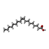
| Macromolecule | Name: PALMITIC ACID / type: ligand / ID: 3 / Number of copies: 3 / Formula: PLM |
|---|---|
| Molecular weight | Theoretical: 256.424 Da |
| Chemical component information |  ChemComp-PLM: |
-Macromolecule #4: 2-acetamido-2-deoxy-beta-D-glucopyranose
| Macromolecule | Name: 2-acetamido-2-deoxy-beta-D-glucopyranose / type: ligand / ID: 4 / Number of copies: 33 / Formula: NAG |
|---|---|
| Molecular weight | Theoretical: 221.208 Da |
| Chemical component information |  ChemComp-NAG: |
-Macromolecule #5: OLEIC ACID
| Macromolecule | Name: OLEIC ACID / type: ligand / ID: 5 / Number of copies: 3 / Formula: OLA |
|---|---|
| Molecular weight | Theoretical: 282.461 Da |
| Chemical component information |  ChemComp-OLA: |
-Experimental details
-Structure determination
| Method | cryo EM |
|---|---|
 Processing Processing | single particle reconstruction |
| Aggregation state | particle |
- Sample preparation
| Buffer | pH: 8 |
|---|---|
| Vitrification | Cryogen name: ETHANE |
- Electron microscopy
| Microscope | FEI TALOS ARCTICA |
|---|---|
| Image recording | Film or detector model: GATAN K3 BIOQUANTUM (6k x 4k) / Average electron dose: 50.0 e/Å2 |
| Electron beam | Acceleration voltage: 300 kV / Electron source:  FIELD EMISSION GUN FIELD EMISSION GUN |
| Electron optics | Illumination mode: FLOOD BEAM / Imaging mode: BRIGHT FIELD / Nominal defocus max: 2.2 µm / Nominal defocus min: 1.2 µm |
| Sample stage | Specimen holder model: FEI TITAN KRIOS AUTOGRID HOLDER / Cooling holder cryogen: NITROGEN |
| Experimental equipment |  Model: Talos Arctica / Image courtesy: FEI Company |
 Movie
Movie Controller


 Z (Sec.)
 Y (Row.)
 X (Col.)


