 Movie
Movie Controller

[English] 日本語
 Yorodumi
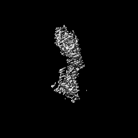
- EMDB-62274: CryoEM structure of Calcineurin-fusion Human endothelin receptor ... -
+ Open data

- Basic information
| Entry |  | |||||||||
|---|---|---|---|---|---|---|---|---|---|---|
| Title | CryoEM structure of Calcineurin-fusion Human endothelin receptor type-B in complex with RES-701-3 | |||||||||
 Map data Map data | ||||||||||
 Sample Sample |
| |||||||||
 Keywords Keywords | GPCR / MEMBRANE PROTEIN | |||||||||
| Function / homology |  Function and homology information Function and homology information: / negative regulation of calcium-mediated signaling / negative regulation of insulin secretion involved in cellular response to glucose stimulus / neuronal action potential propagation / insulin secretion involved in cellular response to glucose stimulus / negative regulation of release of sequestered calcium ion into cytosol / response to redox state / negative regulation of heart rate / 'de novo' protein folding / FK506 binding ...: / negative regulation of calcium-mediated signaling / negative regulation of insulin secretion involved in cellular response to glucose stimulus / neuronal action potential propagation / insulin secretion involved in cellular response to glucose stimulus / negative regulation of release of sequestered calcium ion into cytosol / response to redox state / negative regulation of heart rate / 'de novo' protein folding / FK506 binding / smooth muscle contraction / T cell proliferation / calcium channel inhibitor activity / regulation of release of sequestered calcium ion into cytosol by sarcoplasmic reticulum / Ion homeostasis / release of sequestered calcium ion into cytosol / regulation of cardiac muscle contraction by regulation of the release of sequestered calcium ion / sarcoplasmic reticulum membrane / calcium channel complex / peptidylprolyl isomerase / peptidyl-prolyl cis-trans isomerase activity / calcium channel regulator activity / calcium-mediated signaling / protein maturation / Z disc / Stimuli-sensing channels / protein refolding / positive regulation of cytosolic calcium ion concentration / transmembrane transporter binding / signaling receptor binding / membrane / cytoplasm Similarity search - Function | |||||||||
| Biological species |  Homo sapiens (human) / Homo sapiens (human) /  Streptomyces venezuelae (bacteria) Streptomyces venezuelae (bacteria) | |||||||||
| Method | single particle reconstruction / cryo EM / Resolution: 3.3 Å | |||||||||
 Authors Authors | Shihoya W / Akasaka H / Nureki O | |||||||||
| Funding support |  Japan, 1 items Japan, 1 items
| |||||||||
 Citation Citation | Journal: Nat Commun / Year: 2025 Title: Structure of a lasso peptide bound ET receptor provides insights into the mechanism of GPCR inverse agonism. Authors: Wataru Shihoya / Hiroaki Akasaka / Peter A Jordan / Anna Lechner / Bethany K Okada / Gabriella Costa Machado da Cruz / Fumiya K Sano / Tatsuki Tanaka / Ryo Kawahara / Rajan Chaudhari / ...Authors: Wataru Shihoya / Hiroaki Akasaka / Peter A Jordan / Anna Lechner / Bethany K Okada / Gabriella Costa Machado da Cruz / Fumiya K Sano / Tatsuki Tanaka / Ryo Kawahara / Rajan Chaudhari / Hiroko Masamune / Mark J Burk / Osamu Nureki /  Abstract: Lasso peptides exhibit a unique lariat-like knotted structure imparting exceptional stability and thus show promise as therapeutic agents that target cell-surface receptors. One such receptor is the ...Lasso peptides exhibit a unique lariat-like knotted structure imparting exceptional stability and thus show promise as therapeutic agents that target cell-surface receptors. One such receptor is the human endothelin type B receptor (ET), which is implicated in challenging cancers with poor immunotherapy responsiveness. The Streptomyces-derived lasso peptide, RES-701-3, is a selective inhibitor for ET and a compelling candidate for therapeutic development. However, meager production from a genetically recalcitrant host has limited further structure-activity relationship studies of this potent inhibitor. Here, we report cryo-electron microscopy structures of ET receptor in both its apo form and complex with RES-701-3, facilitated by a calcineurin-fusion strategy. Hydrophobic interactions between RES-701-3 and the transmembrane region of the receptor, especially involving two tryptophan residues, play a crucial role in RES-701-3 binding. Furthermore, RES-701-3 prevents conformational changes associated with G-protein coupling, explaining its inverse agonist activity. A comparative analysis with other lasso peptides and their target proteins highlights the potential of lasso peptides as precise drug candidates for G-protein-coupled receptors. This structural insight into RES-701-3 binding to ET receptor offers valuable information for the development of novel therapeutics targeting this receptor and provides a broader understanding of lasso peptide interactions with human cell-surface receptors. | |||||||||
| History |
|
- Structure visualization
| Supplemental images |
|---|

- Downloads & links
-EMDB archive
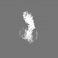
| Map data | emd_62274.map.gz | 26.8 MB | EMDB map data format | |
|---|---|---|---|---|
| Header (meta data) | emd-62274-v30.xmlemd-62274.xml | 19 KB 19 KB | Display Display | EMDB header |
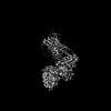
| Images |  emd_62274.png emd_62274.png | 47.4 KB | ||
| Filedesc metadata | emd-62274.cif.gz | 6.8 KB | ||
| Archive directory |  http://ftp.pdbj.org/pub/emdb/structures/EMD-62274ftp://ftp.pdbj.org/pub/emdb/structures/EMD-62274 http://ftp.pdbj.org/pub/emdb/structures/EMD-62274ftp://ftp.pdbj.org/pub/emdb/structures/EMD-62274 | HTTPS FTP |
-Related structure data
| Related structure data | 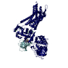 9kdfMC 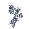 9kdgC C: citing same article ( M: atomic model generated by this map |
|---|---|
| Similar structure data |

-Links
| EMDB pages | EMDB (EBI/PDBe) / EMDataResource |
|---|---|
| Related items in Molecule of the Month |


-Map
| File | Download / File: emd_62274.map.gz / Format: CCP4 / Size: 30.5 MB / Type: IMAGE STORED AS FLOATING POINT NUMBER (4 BYTES) | ||||||||||||||||||||||||||||||||||||
|---|---|---|---|---|---|---|---|---|---|---|---|---|---|---|---|---|---|---|---|---|---|---|---|---|---|---|---|---|---|---|---|---|---|---|---|---|---|
| Projections & slices | Image control
Images are generated by Spider. | ||||||||||||||||||||||||||||||||||||
| Voxel size | X=Y=Z: 1.162 Å | ||||||||||||||||||||||||||||||||||||
| Density |
| ||||||||||||||||||||||||||||||||||||
| Symmetry | Space group: 1 | ||||||||||||||||||||||||||||||||||||
| Details | EMDB XML:
|
 Z (Sec.)
 Y (Row.)
 X (Col.)


-Supplemental data
- Sample components
+Entire : Calcineurin-fusion Human endothelin receptor type-B in complex wi...
+Supramolecule #1: Calcineurin-fusion Human endothelin receptor type-B in complex wi...
+Supramolecule #2: Calcineurin-fusion endothelin receptor type-B
+Supramolecule #3: FKBP16
+Supramolecule #4: RES-701-3
+Supramolecule #5: Endothelin receptor type-B
+Supramolecule #6: Calcineurin
+Macromolecule #1: Calcineurin-fusion endothelin receptor type-B

 Spodoptera frugiperda (fall armyworm)
+Macromolecule #2: Peptidyl-prolyl cis-trans isomerase FKBP1B
+Macromolecule #3: RES-701-3
+Macromolecule #4: CALCIUM ION
+Macromolecule #5: FE (III) ION
+Macromolecule #6: ZINC ION
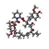
+Macromolecule #7: 8-DEETHYL-8-[BUT-3-ENYL]-ASCOMYCIN
-Experimental details
-Structure determination
| Method | cryo EM |
|---|---|
 Processing Processing | single particle reconstruction |
| Aggregation state | particle |
-Sample preparation
| Buffer | pH: 8 |
|---|---|
| Vitrification | Cryogen name: ETHANE |
- Electron microscopy
| Microscope | TFS KRIOS |
|---|---|
| Image recording | Film or detector model: GATAN K3 (6k x 4k) / Average electron dose: 50.0 e/Å2 |
| Electron beam | Acceleration voltage: 300 kV / Electron source:  FIELD EMISSION GUN FIELD EMISSION GUN |
| Electron optics | Illumination mode: FLOOD BEAM / Imaging mode: BRIGHT FIELD / Nominal defocus max: 1.6 µm / Nominal defocus min: 0.8 µm |
| Experimental equipment |  Model: Titan Krios / Image courtesy: FEI Company |
