 Movie
Movie Controller


+ Open data

- Basic information
| Entry | Database: EMDB / ID: EMD-6226 | |||||||||
|---|---|---|---|---|---|---|---|---|---|---|
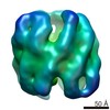
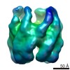
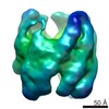
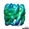
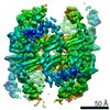
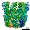
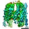
| Title | Substrate-free TRiC | |||||||||
 Map data Map data | substrate-free TRiC (sub-class of AML1-175 + TRiC) | |||||||||
 Sample Sample |
| |||||||||
 Keywords Keywords | eukaryotic chaperonin | |||||||||
| Biological species |  Homo sapiens (human) Homo sapiens (human) | |||||||||
| Method | single particle reconstruction / cryo EM / Resolution: 25.0 Å | |||||||||
 Authors Authors | Roh SH / Kasembeli M / Montoya JG / Trnka M / Lau WC / Burlingame A / Chiu W / Tweardy DJ | |||||||||
 Citation Citation | Journal: Biophys J / Year: 2016 Title: Chaperonin TRiC/CCT Recognizes Fusion Oncoprotein AML1-ETO through Subunit-Specific Interactions. Authors: Soung-Hun Roh / Moses M Kasembeli / Jesús G Galaz-Montoya / Wah Chiu / David J Tweardy /  Abstract: AML1-ETO is the translational product of a chimeric gene created by the stable chromosome translocation t (8;21)(q22;q22). It causes acute myeloid leukemia (AML) by dysregulating the expression of ...AML1-ETO is the translational product of a chimeric gene created by the stable chromosome translocation t (8;21)(q22;q22). It causes acute myeloid leukemia (AML) by dysregulating the expression of genes critical for myeloid cell development and differentiation and recently has been reported to bind multiple subunits of the mammalian cytosolic chaperonin TRiC (or CCT), primarily through its DNA binding domain (AML1-175). Through these interactions, TRiC plays an important role in the synthesis, folding, and activity of AML1-ETO. Using single-particle cryo-electron microscopy, we demonstrate here that a folding intermediate of AML1-ETO's DNA-binding domain (AML1-175) forms a stable complex with apo-TRiC. Our structure reveals that AML1-175 associates directly with a specific subset of TRiC subunits in the open conformation. | |||||||||
| History |
|
- Structure visualization
| Movie |
 Movie viewer Movie viewer |
|---|---|
| Structure viewer | EM map: SurfViewMolmilJmol/JSmol |
| Supplemental images |

- Downloads & links
-EMDB archive
| Map data | emd_6226.map.gz | 58.6 MB | EMDB map data format | |
|---|---|---|---|---|
| Header (meta data) | emd-6226-v30.xmlemd-6226.xml | 8.7 KB 8.7 KB | Display Display | EMDB header |
| Images |  400_6226.gif 400_6226.gif 80_6226.gif 80_6226.gif | 38.8 KB 3.7 KB | ||
| Archive directory |  http://ftp.pdbj.org/pub/emdb/structures/EMD-6226ftp://ftp.pdbj.org/pub/emdb/structures/EMD-6226 http://ftp.pdbj.org/pub/emdb/structures/EMD-6226ftp://ftp.pdbj.org/pub/emdb/structures/EMD-6226 | HTTPS FTP |
-Related structure data
-Links
| EMDB pages | EMDB (EBI/PDBe) / EMDataResource |
|---|
-Map
| File | Download / File: emd_6226.map.gz / Format: CCP4 / Size: 62.5 MB / Type: IMAGE STORED AS FLOATING POINT NUMBER (4 BYTES) | ||||||||||||||||||||||||||||||||||||||||||||||||||||||||||||
|---|---|---|---|---|---|---|---|---|---|---|---|---|---|---|---|---|---|---|---|---|---|---|---|---|---|---|---|---|---|---|---|---|---|---|---|---|---|---|---|---|---|---|---|---|---|---|---|---|---|---|---|---|---|---|---|---|---|---|---|---|---|
| Annotation | substrate-free TRiC (sub-class of AML1-175 + TRiC) | ||||||||||||||||||||||||||||||||||||||||||||||||||||||||||||
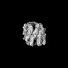
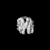
| Projections & slices | Image control
Images are generated by Spider. | ||||||||||||||||||||||||||||||||||||||||||||||||||||||||||||
| Voxel size | X=Y=Z: 2.17 Å | ||||||||||||||||||||||||||||||||||||||||||||||||||||||||||||
| Density |
| ||||||||||||||||||||||||||||||||||||||||||||||||||||||||||||
| Symmetry | Space group: 1 | ||||||||||||||||||||||||||||||||||||||||||||||||||||||||||||
| Details | EMDB XML:
CCP4 map header:
| ||||||||||||||||||||||||||||||||||||||||||||||||||||||||||||
 Z (Sec.)
 Y (Row.)
 X (Col.)


-Supplemental data
- Sample components
-Entire : TRiC
| Entire | Name: TRiC |
|---|---|
| Components |
|
-Supramolecule #1000: TRiC
| Supramolecule | Name: TRiC / type: sample / ID: 1000 / Oligomeric state: 16 / Number unique components: 1 |
|---|---|
| Molecular weight | Theoretical: 1 MDa |
-Macromolecule #1: TCP-1 Ring Complex
| Macromolecule | Name: TCP-1 Ring Complex / type: protein_or_peptide / ID: 1 / Name.synonym: TRiC, CCT / Oligomeric state: 16 / Recombinant expression: No / Database: NCBI |
|---|---|
| Source (natural) | Organism: Homo sapiens (human) / synonym: Human / Cell: HeLa |
| Molecular weight | Theoretical: 1 MDa |
-Experimental details
-Structure determination
| Method | cryo EM |
|---|---|
 Processing Processing | single particle reconstruction |
| Aggregation state | particle |
-Sample preparation
| Concentration | 0.5 mg/mL |
|---|---|
| Buffer | pH: 7.4 Details: 20 mM HEPES, pH 7.4, 5 mM MgCl2, 0.1 mM EDTA, 1 mM DTT, 2% glycerol, 1% PEG8000, 300 mM NaCl, 0.05% OG |
| Grid | Details: 200 mesh Quantifoil holey carbon grid (1.2/1.3), acetone-washed and glow-discharged |
| Vitrification | Cryogen name: ETHANE / Chamber humidity: 80 % / Instrument: LEICA EM GP / Method: Blot for 10 seconds from copper side of grid. |
- Electron microscopy
| Microscope | JEOL 2010F |
|---|---|
| Date | Jan 24, 2014 |
| Image recording | Category: CCD / Film or detector model: GATAN ULTRASCAN 4000 (4k x 4k) / Digitization - Sampling interval: 2.17 µm / Number real images: 263 / Average electron dose: 25 e/Å2 |
| Electron beam | Acceleration voltage: 200 kV / Electron source:  FIELD EMISSION GUN FIELD EMISSION GUN |
| Electron optics | Calibrated magnification: 69124 / Illumination mode: FLOOD BEAM / Imaging mode: BRIGHT FIELD / Cs: 2 mm / Nominal defocus max: 4.0 µm / Nominal defocus min: 1.5 µm / Nominal magnification: 50000 |
| Sample stage | Specimen holder model: GATAN LIQUID NITROGEN |
-Image processing
| CTF correction | Details: particle-based |
|---|---|
| Final reconstruction | Resolution.type: BY AUTHOR / Resolution: 25.0 Å / Resolution method: OTHER / Software - Name: EMAN2, Relion1.2 / Number images used: 11449 |
