Movie
Movie Controller

[English] 日本語
 Yorodumi
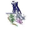
- EMDB-61913: Focused map of Human Lysophosphatidic Acid Receptor 1-Gi complex ... -
+ Open data

- Basic information
| Entry |  | |||||||||
|---|---|---|---|---|---|---|---|---|---|---|
| Title | Focused map of Human Lysophosphatidic Acid Receptor 1-Gi complex bound to CpY | |||||||||
 Map data Map data | ||||||||||
 Sample Sample |
| |||||||||
 Keywords Keywords | GPCR / MEMBRANE PROTEIN | |||||||||
| Biological species |   Homo sapiens (human) / Homo sapiens (human) / | |||||||||
| Method | single particle reconstruction / cryo EM / Resolution: 3.12 Å | |||||||||
 Authors Authors | Akasaka H / Shihoya W / Nureki O | |||||||||
| Funding support |  Japan, 1 items Japan, 1 items
| |||||||||
 Citation Citation | Journal: Commun Biol / Year: 2024 Title: Structural mechanisms of potent lysophosphatidic acid receptor 1 activation by nonlipid basic agonists. Authors: Hiroaki Akasaka / Fumiya K Sano / Wataru Shihoya / Osamu Nureki / Abstract: Lysophosphatidic acid receptor 1 (LPA) is one of the G protein-coupled receptors activated by the lipid mediator, lysophosphatidic acid (LPA). LPA is associated with a variety of diseases, and LPA ...Lysophosphatidic acid receptor 1 (LPA) is one of the G protein-coupled receptors activated by the lipid mediator, lysophosphatidic acid (LPA). LPA is associated with a variety of diseases, and LPA agonists have potential therapeutic value for treating obesity and depression. Although potent nonlipid LPA agonists have recently been identified, the mechanisms of nonlipid molecule-mediated LPA activation remain unclear. Here, we report a cryo-electron microscopy structure of the human LPA-G complex bound to a nonlipid basic agonist, CpY, which has 30-fold higher agonistic activity as compared with LPA. Structural comparisons of LPA with other lipid GPCRs revealed that the negative charge in the characteristic binding pocket of LPA allows the selective recognition of CpY, which lacks a polar head. In addition, our structure show that the ethyl group of CpY directly pushes W271 to fix the active conformation. Endogenous LPA lacks these chemical features, which thus represent the crucial elements of nonlipid agonists that potently activate LPA. This study provides detailed mechanistic insights into the ligand recognition and activation of LPA by nonlipid agonists, expanding the scope for drug development targeting the LPA receptors. | |||||||||
| History |
|
- Structure visualization
| Supplemental images |
|---|

- Downloads & links
-EMDB archive
| Map data | emd_61913.map.gz | 28.8 MB |  EMDB map data format EMDB map data format | |
|---|---|---|---|---|
| Header (meta data) | emd-61913-v30.xmlemd-61913.xml | 15 KB 15 KB | Display Display | EMDB header |
| FSC (resolution estimation) | emd_61913_fsc.xml | 6.6 KB | Display | FSC data file |
| Images |  emd_61913.png emd_61913.png | 35.8 KB | ||
| Masks | emd_61913_msk_1.map | 30.5 MB | Mask map | |
| Filedesc metadata | emd-61913.cif.gz | 4 KB | ||
| Others | emd_61913_half_map_1.map.gzemd_61913_half_map_2.map.gz | 28.4 MB 28.4 MB | ||
| Archive directory |  http://ftp.pdbj.org/pub/emdb/structures/EMD-61913ftp://ftp.pdbj.org/pub/emdb/structures/EMD-61913 http://ftp.pdbj.org/pub/emdb/structures/EMD-61913ftp://ftp.pdbj.org/pub/emdb/structures/EMD-61913 | HTTPS FTP |
-Validation report
| Summary document | emd_61913_validation.pdf.gz | 828.9 KB | Display | EMDB validaton report |
|---|---|---|---|---|
| Full document | emd_61913_full_validation.pdf.gz | 828.5 KB | Display | |
| Data in XML | emd_61913_validation.xml.gz | 13.9 KB | Display | |
| Data in CIF | emd_61913_validation.cif.gz | 17.9 KB | Display | |
| Arichive directory | https://ftp.pdbj.org/pub/emdb/validation_reports/EMD-61913ftp://ftp.pdbj.org/pub/emdb/validation_reports/EMD-61913 | HTTPS FTP |
-Related structure data


-Links
| EMDB pages | EMDB (EBI/PDBe) / EMDataResource |
|---|
-Map
| File | Download / File: emd_61913.map.gz / Format: CCP4 / Size: 30.5 MB / Type: IMAGE STORED AS FLOATING POINT NUMBER (4 BYTES) | ||||||||||||||||||||||||||||||||||||
|---|---|---|---|---|---|---|---|---|---|---|---|---|---|---|---|---|---|---|---|---|---|---|---|---|---|---|---|---|---|---|---|---|---|---|---|---|---|
| Projections & slices | Image control
Images are generated by Spider. | ||||||||||||||||||||||||||||||||||||
| Voxel size | X=Y=Z: 1.162 Å | ||||||||||||||||||||||||||||||||||||
| Density |
| ||||||||||||||||||||||||||||||||||||
| Symmetry | Space group: 1 | ||||||||||||||||||||||||||||||||||||
| Details | EMDB XML:
|
 Z (Sec.)
 Y (Row.)
 X (Col.)


-Supplemental data
-Mask #1
| File | emd_61913_msk_1.map | ||||||||||||
|---|---|---|---|---|---|---|---|---|---|---|---|---|---|
| Projections & Slices |
| ||||||||||||
| Density Histograms |


-Half map: #1
| File | emd_61913_half_map_1.map | ||||||||||||
|---|---|---|---|---|---|---|---|---|---|---|---|---|---|
| Projections & Slices |
| ||||||||||||
| Density Histograms |


-Half map: #2
| File | emd_61913_half_map_2.map | ||||||||||||
|---|---|---|---|---|---|---|---|---|---|---|---|---|---|
| Projections & Slices |
| ||||||||||||
| Density Histograms |


- Sample components
-Entire : Human Lysophosphatidic Acid Receptor 1-Gi complex bound to ONO-0740556
| Entire | Name: Human Lysophosphatidic Acid Receptor 1-Gi complex bound to ONO-0740556 |
|---|---|
| Components |
|
-Supramolecule #1: Human Lysophosphatidic Acid Receptor 1-Gi complex bound to ONO-0740556
| Supramolecule | Name: Human Lysophosphatidic Acid Receptor 1-Gi complex bound to ONO-0740556 type: complex / ID: 1 / Parent: 0 / Macromolecule list: #1-#5 |
|---|
-Supramolecule #2: Guanine nucleotide-binding protein G(I)/G(S)/G(T) subunit beta-1
| Supramolecule | Name: Guanine nucleotide-binding protein G(I)/G(S)/G(T) subunit beta-1 type: complex / ID: 2 / Parent: 1 / Macromolecule list: #1 |
|---|---|
| Source (natural) | Organism: |
-Supramolecule #3: Guanine nucleotide-binding protein G(i) subunit alpha-1
| Supramolecule | Name: Guanine nucleotide-binding protein G(i) subunit alpha-1 type: complex / ID: 3 / Parent: 1 / Macromolecule list: #2 |
|---|---|
| Source (natural) | Organism: Homo sapiens (human) |
-Supramolecule #4: Guanine nucleotide-binding protein G(I)/G(S)/G(O) subunit gamma-2
| Supramolecule | Name: Guanine nucleotide-binding protein G(I)/G(S)/G(O) subunit gamma-2 type: complex / ID: 4 / Parent: 1 / Macromolecule list: #3 |
|---|---|
| Source (natural) | Organism: |
-Supramolecule #5: Lysophosphatidic Acid Receptor 1
| Supramolecule | Name: Lysophosphatidic Acid Receptor 1 / type: complex / ID: 5 / Parent: 1 / Macromolecule list: #4 |
|---|---|
| Source (natural) | Organism: Homo sapiens (human) |
-Supramolecule #6: scFv16
| Supramolecule | Name: scFv16 / type: complex / ID: 6 / Parent: 1 / Macromolecule list: #5 |
|---|---|
| Source (natural) | Organism: |
-Experimental details
-Structure determination
| Method | cryo EM |
|---|---|
 Processing Processing | single particle reconstruction |
| Aggregation state | particle |
-Sample preparation
| Concentration | 7 mg/mL |
|---|---|
| Buffer | pH: 8 |
| Vitrification | Cryogen name: ETHANE |
- Electron microscopy
| Microscope | TFS KRIOS |
|---|---|
| Image recording | Film or detector model: GATAN K3 (6k x 4k) / Average electron dose: 50.0 e/Å2 |
| Electron beam | Acceleration voltage: 300 kV / Electron source:  FIELD EMISSION GUN FIELD EMISSION GUN |
| Electron optics | Illumination mode: FLOOD BEAM / Imaging mode: BRIGHT FIELD / Nominal defocus max: 2.5 µm / Nominal defocus min: 0.5 µm |
| Experimental equipment |  Model: Titan Krios / Image courtesy: FEI Company |