[English] 日本語
 Yorodumi
- EMDB-61814: Human glucose 6 phosphate catalytic subunit 1 (hG6PC1) bound with F6P. -
+ Open data
- Basic information
| Entry |  | |||||||||
|---|---|---|---|---|---|---|---|---|---|---|
| Title | Human glucose 6 phosphate catalytic subunit 1 (hG6PC1) bound with F6P. | |||||||||
 Map data Map data | ||||||||||
 Sample Sample |
| |||||||||
 Keywords Keywords | membrane protein / glucose metabolism / HYDROLASE | |||||||||
| Function / homology |  Function and homology information Function and homology informationglucose-6-phosphatase / Glycogen storage disease type Ia (G6PC) / glucose-6-phosphatase activity / phosphotransferase activity, alcohol group as acceptor / glucose-6-phosphate transport / response to resveratrol / urate metabolic process / glucose 6-phosphate metabolic process / response to carbohydrate / Gluconeogenesis ...glucose-6-phosphatase / Glycogen storage disease type Ia (G6PC) / glucose-6-phosphatase activity / phosphotransferase activity, alcohol group as acceptor / glucose-6-phosphate transport / response to resveratrol / urate metabolic process / glucose 6-phosphate metabolic process / response to carbohydrate / Gluconeogenesis / glycogen catabolic process / triglyceride metabolic process / response to food / glycogen metabolic process / phosphate ion binding / steroid metabolic process / FOXO-mediated transcription of oxidative stress, metabolic and neuronal genes / cholesterol homeostasis / gluconeogenesis / multicellular organism growth / cellular response to insulin stimulus / glucose homeostasis / regulation of gene expression / endoplasmic reticulum membrane / membrane Similarity search - Function | |||||||||
| Biological species |  Homo sapiens (human) Homo sapiens (human) | |||||||||
| Method | single particle reconstruction / cryo EM / Resolution: 3.3 Å | |||||||||
 Authors Authors | Chen Q / Zhao Y / Wang Y | |||||||||
| Funding support |  China, 1 items China, 1 items
| |||||||||
 Citation Citation |  Journal: Cell Discov / Year: 2025 Journal: Cell Discov / Year: 2025Title: The induced-fit and catalytic mechanisms of human G6PC1. Authors: Qihao Chen / Yuhang Wang / Renjie Li / Qinru Bai / Yan Zhao /  Abstract: Human glucose-6-phosphatase catalytic subunit 1 (hG6PC1) is a key enzyme in glucose metabolism, governing the final common step of gluconeogenesis and glycogenolysis, and directly regulating energy ...Human glucose-6-phosphatase catalytic subunit 1 (hG6PC1) is a key enzyme in glucose metabolism, governing the final common step of gluconeogenesis and glycogenolysis, and directly regulating energy homeostasis. Aberrant mutations in G6PC1 directly cause glycogen storage disease type 1a, which is characterized by chronic hypoglycemia and glycogen accumulation. Additionally, abnormal G6PC1 function leads to increased fasting blood glucose. Consequently, it is a critical target for treating glucose metabolism disorders. In this study, we determine the cryo-EM structures of G6PC1 in both the partially open and fully open states, in either the apo form or in complex with the substrates G6P or F6P and the product phosphate. These structures offer distinct insights into the mechanism of hydrolysis and induced-fit, providing a structural foundation for the diagnostic analysis of disease-causing mutations in G6PC1. Moreover, we propose a potential mechanism by which phosphatidylserine regulates G6PC1 activity, providing a novel perspective on its role and implications. | |||||||||
| History |
|
- Structure visualization
| Supplemental images |
|---|
- Downloads & links
-EMDB archive
| Map data |  emd_61814.map.gz emd_61814.map.gz | 59.7 MB |  EMDB map data format EMDB map data format | |
|---|---|---|---|---|
| Header (meta data) |  emd-61814-v30.xml emd-61814-v30.xml emd-61814.xml emd-61814.xml | 14.7 KB 14.7 KB | Display Display |  EMDB header EMDB header |
| Images |  emd_61814.png emd_61814.png | 90 KB | ||
| Filedesc metadata |  emd-61814.cif.gz emd-61814.cif.gz | 5.9 KB | ||
| Others |  emd_61814_half_map_1.map.gz emd_61814_half_map_1.map.gz emd_61814_half_map_2.map.gz emd_61814_half_map_2.map.gz | 59.3 MB 59.3 MB | ||
| Archive directory |  http://ftp.pdbj.org/pub/emdb/structures/EMD-61814 http://ftp.pdbj.org/pub/emdb/structures/EMD-61814 ftp://ftp.pdbj.org/pub/emdb/structures/EMD-61814 ftp://ftp.pdbj.org/pub/emdb/structures/EMD-61814 | HTTPS FTP |
-Validation report
| Summary document |  emd_61814_validation.pdf.gz emd_61814_validation.pdf.gz | 1.1 MB | Display |  EMDB validaton report EMDB validaton report |
|---|---|---|---|---|
| Full document |  emd_61814_full_validation.pdf.gz emd_61814_full_validation.pdf.gz | 1.1 MB | Display | |
| Data in XML |  emd_61814_validation.xml.gz emd_61814_validation.xml.gz | 12.3 KB | Display | |
| Data in CIF |  emd_61814_validation.cif.gz emd_61814_validation.cif.gz | 14.5 KB | Display | |
| Arichive directory |  https://ftp.pdbj.org/pub/emdb/validation_reports/EMD-61814 https://ftp.pdbj.org/pub/emdb/validation_reports/EMD-61814 ftp://ftp.pdbj.org/pub/emdb/validation_reports/EMD-61814 ftp://ftp.pdbj.org/pub/emdb/validation_reports/EMD-61814 | HTTPS FTP |
-Related structure data
| Related structure data |  9jtoMC  9jtlC  9jtmC  9jtnC M: atomic model generated by this map C: citing same article ( |
|---|---|
| Similar structure data | Similarity search - Function & homology  F&H Search F&H Search |
- Links
Links
| EMDB pages |  EMDB (EBI/PDBe) / EMDB (EBI/PDBe) /  EMDataResource EMDataResource |
|---|---|
| Related items in Molecule of the Month |
- Map
Map
| File |  Download / File: emd_61814.map.gz / Format: CCP4 / Size: 64 MB / Type: IMAGE STORED AS FLOATING POINT NUMBER (4 BYTES) Download / File: emd_61814.map.gz / Format: CCP4 / Size: 64 MB / Type: IMAGE STORED AS FLOATING POINT NUMBER (4 BYTES) | ||||||||||||||||||||||||||||||||||||
|---|---|---|---|---|---|---|---|---|---|---|---|---|---|---|---|---|---|---|---|---|---|---|---|---|---|---|---|---|---|---|---|---|---|---|---|---|---|
| Projections & slices | Image control
Images are generated by Spider. | ||||||||||||||||||||||||||||||||||||
| Voxel size | X=Y=Z: 0.85 Å | ||||||||||||||||||||||||||||||||||||
| Density |
| ||||||||||||||||||||||||||||||||||||
| Symmetry | Space group: 1 | ||||||||||||||||||||||||||||||||||||
| Details | EMDB XML:
|
-Supplemental data
-Half map: #2
| File | emd_61814_half_map_1.map | ||||||||||||
|---|---|---|---|---|---|---|---|---|---|---|---|---|---|
| Projections & Slices |
| ||||||||||||
| Density Histograms |
-Half map: #1
| File | emd_61814_half_map_2.map | ||||||||||||
|---|---|---|---|---|---|---|---|---|---|---|---|---|---|
| Projections & Slices |
| ||||||||||||
| Density Histograms |
- Sample components
-Entire : Glucose 6 phosphate catalytic subunit 1
| Entire | Name: Glucose 6 phosphate catalytic subunit 1 |
|---|---|
| Components |
|
-Supramolecule #1: Glucose 6 phosphate catalytic subunit 1
| Supramolecule | Name: Glucose 6 phosphate catalytic subunit 1 / type: complex / ID: 1 / Parent: 0 / Macromolecule list: #1 |
|---|---|
| Source (natural) | Organism:  Homo sapiens (human) Homo sapiens (human) |
-Macromolecule #1: Glucose-6-phosphatase catalytic subunit 1,GSlinker-HRV3C-GFP-twin...
| Macromolecule | Name: Glucose-6-phosphatase catalytic subunit 1,GSlinker-HRV3C-GFP-twin strep type: protein_or_peptide / ID: 1 / Number of copies: 1 / Enantiomer: LEVO / EC number: glucose-6-phosphatase |
|---|---|
| Source (natural) | Organism:  Homo sapiens (human) Homo sapiens (human) |
| Molecular weight | Theoretical: 71.877039 KDa |
| Recombinant expression | Organism:  Homo sapiens (human) Homo sapiens (human) |
| Sequence | String: MEEGMNVLHD FGIQSTHYLQ VNYQDSQDWF ILVSVIADLR NAFYVLFPIW FHLQEAVGIK LLWVAVIGDW LNLVFKWILF GQRPYWWVL DTDYYSNTSV PLIKQFPVTC ETGPGSPSGH AMGTAGVYYV MVTSTLSIFQ GKIKPTYRFR CLNVILWLGF W AVQLNVCL ...String: MEEGMNVLHD FGIQSTHYLQ VNYQDSQDWF ILVSVIADLR NAFYVLFPIW FHLQEAVGIK LLWVAVIGDW LNLVFKWILF GQRPYWWVL DTDYYSNTSV PLIKQFPVTC ETGPGSPSGH AMGTAGVYYV MVTSTLSIFQ GKIKPTYRFR CLNVILWLGF W AVQLNVCL SRIYLAANFP HQVVAGVLSG IAVAETFSHI HSIYNASLKK YFLITFFLFS FAIGFYLLLK GLGVDLLWTL EK AQRWCEQ PEWVHIDTTP FASLLKNLGT LFGLGLALNS SMYRESCKGK LSKWLPFRLS SIVASLVLLH VFDSLKPPSQ VEL VFYVLS FCKSAVVPLA SVSVIPYCLA QVLGQPHKKS LGGSGGGSGG GSGGGSGGGA AALEVLFQGP SKGEELFTGV VPIL VELDG DVNGHKFSVR GEGEGDATNG KLTLKFICTT GKLPVPWPTL VTTLTYGVQC FSRYPDHMKR HDFFKSAMPE GYVQE RTIS FKDDGTYKTR AEVKFEGDTL VNRIELKGID FKEDGNILGH KLEYNFNSHN VYITADKQKN GIKANFKIRH NVEDGS VQL ADHYQQNTPI GDGPVLLPDN HYLSTQSVLS KDPNEKRDHM VLLEFVTAAG ITHGMDEWSH PQFEKGGGSG GGSGGSA WS HPQFEK UniProtKB: Glucose-6-phosphatase catalytic subunit 1 |
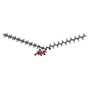
-Macromolecule #2: O-[(R)-{[(2R)-2,3-bis(octadecanoyloxy)propyl]oxy}(hydroxy)phospho...
| Macromolecule | Name: O-[(R)-{[(2R)-2,3-bis(octadecanoyloxy)propyl]oxy}(hydroxy)phosphoryl]-L-serine type: ligand / ID: 2 / Number of copies: 1 / Formula: P5S |
|---|---|
| Molecular weight | Theoretical: 792.075 Da |
| Chemical component information |  ChemComp-P5S: |
-Macromolecule #3: 6-O-phosphono-beta-D-fructofuranose
| Macromolecule | Name: 6-O-phosphono-beta-D-fructofuranose / type: ligand / ID: 3 / Number of copies: 1 / Formula: F6P |
|---|---|
| Molecular weight | Theoretical: 260.136 Da |
| Chemical component information |  ChemComp-F6P: |
-Experimental details
-Structure determination
| Method | cryo EM |
|---|---|
 Processing Processing | single particle reconstruction |
| Aggregation state | particle |
- Sample preparation
| Buffer | pH: 8 |
|---|---|
| Vitrification | Cryogen name: ETHANE |
- Electron microscopy
| Microscope | FEI TALOS ARCTICA |
|---|---|
| Image recording | Film or detector model: GATAN K3 BIOCONTINUUM (6k x 4k) / Average electron dose: 60.0 e/Å2 |
| Electron beam | Acceleration voltage: 300 kV / Electron source:  FIELD EMISSION GUN FIELD EMISSION GUN |
| Electron optics | Illumination mode: FLOOD BEAM / Imaging mode: BRIGHT FIELD / Nominal defocus max: 2.2 µm / Nominal defocus min: 1.2 µm |
| Experimental equipment |  Model: Talos Arctica / Image courtesy: FEI Company |
 Movie
Movie Controller


 Z (Sec.)
 Y (Row.)
 X (Col.)


