 Movie
Movie Controller


+ Open data

- Basic information
| Entry |  | |||||||||
|---|---|---|---|---|---|---|---|---|---|---|
| Title | guide-bound NbaSPARDA complexes | |||||||||
 Map data Map data | ||||||||||
 Sample Sample |
| |||||||||
 Keywords Keywords | protein / RNA / RNA BINDING PROTEIN/RNA / RNA BINDING PROTEIN-RNA complex | |||||||||
| Biological species |  Novosphingopyxis baekryungensis DSM 16222 (bacteria) Novosphingopyxis baekryungensis DSM 16222 (bacteria) | |||||||||
| Method | single particle reconstruction / cryo EM / Resolution: 2.93 Å | |||||||||
 Authors Authors | Zhuang L | |||||||||
| Funding support | 1 items
| |||||||||
 Citation Citation | Journal: Cell Res / Year: 2025 Title: Target DNA-induced filament formation and nuclease activation of SPARDA complex. Authors: Feng Wang / Haijiang Xu / Chendi Zhang / Jialin Xue / Zhuang Li /  Abstract: The short Argonaute-based bacterial defense system, SPARDA (Short Prokaryotic Argonaute and DNase/RNase-APAZ), utilizes guide RNA to target invading complementary DNA and exhibits collateral nuclease ...The short Argonaute-based bacterial defense system, SPARDA (Short Prokaryotic Argonaute and DNase/RNase-APAZ), utilizes guide RNA to target invading complementary DNA and exhibits collateral nuclease activity, leading to cell death or dormancy. However, its detailed mechanisms remain poorly understood. In this study, we investigated the SPARDA system from Novosphingopyxis baekryungensis (NbaSPARDA) and discovered an unexpected filament configuration upon target DNA binding, which strongly correlated with collateral nuclease activity. Filament formation and nuclease activation require a guide-target heteroduplex of sufficient length with perfect complementarity at the central region. A series of cryo-EM structures of NbaSPARDA complexes, loaded with guide RNA, target DNA of varying lengths, and substrate ssDNA, were determined at ~3.0 Å resolution. Structural analyses indicated that guide RNA binding induces dimerization of the NbaSPARDA complex, while target DNA engagement disrupts this dimerization. Further propagation of the guide-target heteroduplex triggers filament formation through a checkpoint mechanism. The NbaSPARDA filament consists of a backbone formed by interlocking short Argonaute proteins, with an inner layer composed of DREN nuclease domains. Filament formation leads to tetramerization of the monomeric DREN nuclease domain, activating its collateral nuclease activity against environmental nucleic acids - a feature leveraged for molecular diagnostics. For bacteria heterologously expressing the NbaSPARDA system, defense against invading bacteriophages and plasmids relies on filament formation. Collectively, these findings illustrate the detailed working mechanism of the NbaSPARDA complex and highlight the importance of its filament formation in host defense. | |||||||||
| History |
|
- Structure visualization
| Supplemental images |
|---|

- Downloads & links
-EMDB archive
| Map data | emd_61769.map.gz | 59.7 MB |  EMDB map data format EMDB map data format | |
|---|---|---|---|---|
| Header (meta data) | emd-61769-v30.xmlemd-61769.xml | 18.7 KB 18.7 KB | Display Display | EMDB header |
| FSC (resolution estimation) | emd_61769_fsc.xml | 8.4 KB | Display | FSC data file |
| Images |  emd_61769.png emd_61769.png | 116.6 KB | ||
| Filedesc metadata | emd-61769.cif.gz | 6.4 KB | ||
| Others | emd_61769_half_map_1.map.gzemd_61769_half_map_2.map.gz | 59.2 MB 59.2 MB | ||
| Archive directory |  http://ftp.pdbj.org/pub/emdb/structures/EMD-61769ftp://ftp.pdbj.org/pub/emdb/structures/EMD-61769 http://ftp.pdbj.org/pub/emdb/structures/EMD-61769ftp://ftp.pdbj.org/pub/emdb/structures/EMD-61769 | HTTPS FTP |
-Related structure data
| Related structure data |  9jsbMC 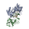 9jspC  9jszC  9jt2C M: atomic model generated by this map C: citing same article ( |
|---|
-Links
| EMDB pages | EMDB (EBI/PDBe) / EMDataResource |
|---|
-Map
| File | Download / File: emd_61769.map.gz / Format: CCP4 / Size: 64 MB / Type: IMAGE STORED AS FLOATING POINT NUMBER (4 BYTES) | ||||||||||||||||||||||||||||||||||||
|---|---|---|---|---|---|---|---|---|---|---|---|---|---|---|---|---|---|---|---|---|---|---|---|---|---|---|---|---|---|---|---|---|---|---|---|---|---|
| Projections & slices | Image control
Images are generated by Spider. | ||||||||||||||||||||||||||||||||||||
| Voxel size | X=Y=Z: 0.85 Å | ||||||||||||||||||||||||||||||||||||
| Density |
| ||||||||||||||||||||||||||||||||||||
| Symmetry | Space group: 1 | ||||||||||||||||||||||||||||||||||||
| Details | EMDB XML:
|
 Z (Sec.)
 Y (Row.)
 X (Col.)


-Supplemental data
-Half map: #2
| File | emd_61769_half_map_1.map | ||||||||||||
|---|---|---|---|---|---|---|---|---|---|---|---|---|---|
| Projections & Slices |
| ||||||||||||
| Density Histograms |


-Half map: #1
| File | emd_61769_half_map_2.map | ||||||||||||
|---|---|---|---|---|---|---|---|---|---|---|---|---|---|
| Projections & Slices |
| ||||||||||||
| Density Histograms |


- Sample components
-Entire : PRO-RNA complex
| Entire | Name: PRO-RNA complex |
|---|---|
| Components |
|
-Supramolecule #1: PRO-RNA complex
| Supramolecule | Name: PRO-RNA complex / type: complex / ID: 1 / Parent: 0 / Macromolecule list: #1-#3 |
|---|---|
| Source (natural) | Organism: Novosphingopyxis baekryungensis DSM 16222 (bacteria) |
-Macromolecule #1: Ago
| Macromolecule | Name: Ago / type: protein_or_peptide / ID: 1 / Number of copies: 2 / Enantiomer: LEVO |
|---|---|
| Source (natural) | Organism: Novosphingopyxis baekryungensis DSM 16222 (bacteria) |
| Molecular weight | Theoretical: 54.492809 KDa |
| Recombinant expression | Organism: |
| Sequence | String: MTFETRIFDE PELEFGDHHH HQDPRLGLSE AGPLQTFLGD VIKIGVVGNS KTIEDTRKFI ETVSSGVEGK GEKHPNMHPP FPGLGNQSP YRCRFEIEDG ATAALTKSKL DKIGKEPDHY RAVEMAVDEI IGELQAMDDG GSRPDVAIIA LPVKLLERVW N AKVDARGT ...String: MTFETRIFDE PELEFGDHHH HQDPRLGLSE AGPLQTFLGD VIKIGVVGNS KTIEDTRKFI ETVSSGVEGK GEKHPNMHPP FPGLGNQSP YRCRFEIEDG ATAALTKSKL DKIGKEPDHY RAVEMAVDEI IGELQAMDDG GSRPDVAIIA LPVKLLERVW N AKVDARGT TEKSDSSGSD APNFRGMLKA KAMGLSFPIQ IVWEDVIDDK VTIPQKVKES SSRKIQDIAG RTWNLMTSLY YK GSGRIPW RRMPLEGEFS ACYVGISFYR EADGQQLFTS AAQMFDERGR GFVLKGRRAR TESRGRHPYM AREDAKKIIE DVL AAYKLH HKTLPARVFI LKTSRFKDEE ADGIIAALDE AGTELRDLVW VQESYTARIL RDGNYPVLRG TFVDLHGKGL LYTS GSMPY YGTYPGKYDP NPLLLCPHHT SESTVAQLAE EIFSLTKVNW NSTQMNQRLP IPIRAARKVG EVLKYVGEGE VISAD YRKY I |
-Macromolecule #2: DREN-APAZ
| Macromolecule | Name: DREN-APAZ / type: protein_or_peptide / ID: 2 / Number of copies: 2 / Enantiomer: LEVO |
|---|---|
| Source (natural) | Organism: Novosphingopyxis baekryungensis DSM 16222 (bacteria) |
| Molecular weight | Theoretical: 50.419645 KDa |
| Recombinant expression | Organism: |
| Sequence | String: MTKKITANQI IGEIGENEVR GRFLTLGWQF DGRSRLEAGI DGIAEVMNEG QPMARMIAVQ IKSTKEGKYT SESDTSFTYL LRTQDLAYW RGSNLPVIVV FYRQSDHSFY WKEVSRDAGP GERRLNIDKV ADLFNASTVN KLAALTVPKT GLGYYVPPLG G GEDALINM ...String: MTKKITANQI IGEIGENEVR GRFLTLGWQF DGRSRLEAGI DGIAEVMNEG QPMARMIAVQ IKSTKEGKYT SESDTSFTYL LRTQDLAYW RGSNLPVIVV FYRQSDHSFY WKEVSRDAGP GERRLNIDKV ADLFNASTVN KLAALTVPKT GLGYYVPPLG G GEDALINM LPLTLPNEMY IASTTYEPRK AIAVILNGDG PKRFDWVING GTFWSFHDPR TSACSEIVDI DQVEAINTKE LA LHDDIDE QNRFSHLLRQ TLRYQTDSDL GWDKDHKALY FRAIEREVSR NFAYTSSKKK TDANVVSVFK NSKDETRVSF VRH HAFSPR FELMADQWYL IITPTYYYTT NGYAPHQFAA PLLAGKKRLD KSAALRGQVI MWHRFLTQSD HEDLFHSEET PEAY LMFGE PPSIHLDVRV PEDGWVKEKV KRIDEAAQGE GLFSDDI |
-Macromolecule #3: RNA (5'-R(P*AP*UP*AP*CP*U)-3')
| Macromolecule | Name: RNA (5'-R(P*AP*UP*AP*CP*U)-3') / type: rna / ID: 3 / Number of copies: 2 |
|---|---|
| Source (natural) | Organism: Novosphingopyxis baekryungensis DSM 16222 (bacteria) |
| Molecular weight | Theoretical: 1.530968 KDa |
| Sequence | String: AUACU |
-Macromolecule #4: MAGNESIUM ION
| Macromolecule | Name: MAGNESIUM ION / type: ligand / ID: 4 / Number of copies: 2 / Formula: MG |
|---|---|
| Molecular weight | Theoretical: 24.305 Da |
-Experimental details
-Structure determination
| Method | cryo EM |
|---|---|
 Processing Processing | single particle reconstruction |
| Aggregation state | particle |
-Sample preparation
| Buffer | pH: 7.5 |
|---|---|
| Vitrification | Cryogen name: ETHANE |
- Electron microscopy
| Microscope | FEI TALOS ARCTICA |
|---|---|
| Image recording | Film or detector model: GATAN K3 BIOQUANTUM (6k x 4k) / Average electron dose: 54.0 e/Å2 |
| Electron beam | Acceleration voltage: 300 kV / Electron source:  FIELD EMISSION GUN FIELD EMISSION GUN |
| Electron optics | Illumination mode: FLOOD BEAM / Imaging mode: BRIGHT FIELD / Nominal defocus max: 2.2 µm / Nominal defocus min: 1.2 µm |
| Experimental equipment |  Model: Talos Arctica / Image courtesy: FEI Company |

