 Movie
Movie Controller


+ Open data

- Basic information
| Entry | 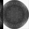 | |||||||||||||||
|---|---|---|---|---|---|---|---|---|---|---|---|---|---|---|---|---|
| Title | Cryo-EM structure of a T=3 VLP of RHDV GI.2 | |||||||||||||||
 Map data Map data | ||||||||||||||||
 Sample Sample |
| |||||||||||||||
 Keywords Keywords | Calicivirus / viral assembly / rabbit hemorrhagic disease virus (RHDV) / major capsid protein / virus-like particle / VIRUS LIKE PARTICLE | |||||||||||||||
| Function / homology | Calicivirus coat protein / Calicivirus coat protein / virion component / Picornavirus/Calicivirus coat protein / Viral coat protein subunit / host cell cytoplasm / Capsid protein Function and homology information Function and homology information | |||||||||||||||
| Biological species |  Rabbit hemorrhagic disease virus 2 Rabbit hemorrhagic disease virus 2 | |||||||||||||||
| Method | single particle reconstruction / cryo EM / Resolution: 2.45 Å | |||||||||||||||
 Authors Authors | Ruan Z / Shao Q / Song Y / Hu B / Fan Z / Wei H / Liu Y / Wang F / Fang Q | |||||||||||||||
| Funding support |  China, 4 items China, 4 items
| |||||||||||||||
 Citation Citation | Journal: J Virol / Year: 2024 Title: Near-atomic structures of RHDV reveal insights into capsid assembly and different conformations between mature virion and VLP. Authors: Zhiyang Ruan / Qianqian Shao / Yanhua Song / Bo Hu / Zhiyu Fan / Houjun Wei / Yunshu Liu / Fang Wang / Qianglin Fang / Abstract: Rabbit hemorrhagic disease virus (RHDV) poses a significant threat to rabbits, causing substantial economic losses in rabbit farming. The virus also endangers wild populations of rabbit species and ...Rabbit hemorrhagic disease virus (RHDV) poses a significant threat to rabbits, causing substantial economic losses in rabbit farming. The virus also endangers wild populations of rabbit species and the predatory animals that rely on rabbits as a food source, thereby disturbing the ecological balance. However, the structural understanding of RHDV has been limited due to the lack of high-resolution structures. Here, we present the first high-resolution cryo-EM structures of the mature virion and virus-like particles (VLPs) derived from both full-length and N-terminal arm (NTA)-truncated VP60. These structures reveal intricate structural details of the icosahedral capsid and crucial NTA-mediated interactions essential for capsid assembly. In addition, dramatic conformational differences are unexpectedly observed between the mature virion and VLP. The protruding spikes of the A-B dimers adopt a "raised" state in the mature virion and a "resting" state in the VLP. These findings enhance our understanding of the structure, assembly, and conformational dynamics of the RHDV capsid, laying the essential groundwork for further virological research and therapeutic advancements.IMPORTANCERHDV is a pathogen with significant economic and ecological impact. By presenting the first high-resolution cryo-EM structures of RHDV, we have uncovered detailed interactions among neighboring VP60 subunits of the icosahedral capsid. The NTA of VP60 is uniquely clustered around the threefold axis of the capsid, probably play a critical role in dragging the six VP60 dimers around the threefold axis during capsid assembly. Additionally, we observed dramatic conformational differences between the mature virion and VLPs. VLPs are commonly used for vaccine development, under the assumption that their structure closely resembles that of the mature virion. Our findings significantly advance the understanding of the RHDV capsid structure, which may be used for developing potential therapeutic strategies against RHDV. | |||||||||||||||
| History |
|
- Structure visualization
| Supplemental images |
|---|

- Downloads & links
-EMDB archive
| Map data | emd_61529.map.gz | 681.9 MB | EMDB map data format | |
|---|---|---|---|---|
| Header (meta data) | emd-61529-v30.xmlemd-61529.xml | 17.2 KB 17.2 KB | Display Display | EMDB header |
| FSC (resolution estimation) | emd_61529_fsc.xml | 20.2 KB | Display | FSC data file |
| Images |  emd_61529.png emd_61529.png | 220.9 KB | ||
| Masks | emd_61529_msk_1.map | 729 MB | Mask map | |
| Filedesc metadata | emd-61529.cif.gz | 6.1 KB | ||
| Others | emd_61529_half_map_1.map.gzemd_61529_half_map_2.map.gz | 592.5 MB 592.6 MB | ||
| Archive directory |  http://ftp.pdbj.org/pub/emdb/structures/EMD-61529ftp://ftp.pdbj.org/pub/emdb/structures/EMD-61529 http://ftp.pdbj.org/pub/emdb/structures/EMD-61529ftp://ftp.pdbj.org/pub/emdb/structures/EMD-61529 | HTTPS FTP |
-Validation report
| Summary document | emd_61529_validation.pdf.gz | 1.2 MB | Display | EMDB validaton report |
|---|---|---|---|---|
| Full document | emd_61529_full_validation.pdf.gz | 1.2 MB | Display | |
| Data in XML | emd_61529_validation.xml.gz | 28.4 KB | Display | |
| Data in CIF | emd_61529_validation.cif.gz | 38.2 KB | Display | |
| Arichive directory | https://ftp.pdbj.org/pub/emdb/validation_reports/EMD-61529ftp://ftp.pdbj.org/pub/emdb/validation_reports/EMD-61529 | HTTPS FTP |
-Related structure data
| Related structure data | 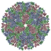 9jjjMC 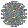 9jjgC 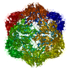 9jjhC 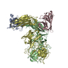 9jjiC M: atomic model generated by this map C: citing same article ( |
|---|---|
| Similar structure data |
-Links
| EMDB pages | EMDB (EBI/PDBe) / EMDataResource |
|---|---|
| Related items in Molecule of the Month |

-Map
| File | Download / File: emd_61529.map.gz / Format: CCP4 / Size: 729 MB / Type: IMAGE STORED AS FLOATING POINT NUMBER (4 BYTES) | ||||||||||||||||||||||||||||||||||||
|---|---|---|---|---|---|---|---|---|---|---|---|---|---|---|---|---|---|---|---|---|---|---|---|---|---|---|---|---|---|---|---|---|---|---|---|---|---|
| Projections & slices | Image control
Images are generated by Spider. | ||||||||||||||||||||||||||||||||||||
| Voxel size | X=Y=Z: 1.074 Å | ||||||||||||||||||||||||||||||||||||
| Density |
| ||||||||||||||||||||||||||||||||||||
| Symmetry | Space group: 1 | ||||||||||||||||||||||||||||||||||||
| Details | EMDB XML:
|
 Z (Sec.)
 Y (Row.)
 X (Col.)


-Supplemental data
-Mask #1
| File | emd_61529_msk_1.map | ||||||||||||
|---|---|---|---|---|---|---|---|---|---|---|---|---|---|
| Projections & Slices |
| ||||||||||||
| Density Histograms |


-Half map: #1
| File | emd_61529_half_map_1.map | ||||||||||||
|---|---|---|---|---|---|---|---|---|---|---|---|---|---|
| Projections & Slices |
| ||||||||||||
| Density Histograms |


-Half map: #2
| File | emd_61529_half_map_2.map | ||||||||||||
|---|---|---|---|---|---|---|---|---|---|---|---|---|---|
| Projections & Slices |
| ||||||||||||
| Density Histograms |


- Sample components
-Entire : Rabbit hemorrhagic disease virus 2
| Entire | Name: Rabbit hemorrhagic disease virus 2 |
|---|---|
| Components |
|
-Supramolecule #1: Rabbit hemorrhagic disease virus 2
| Supramolecule | Name: Rabbit hemorrhagic disease virus 2 / type: virus / ID: 1 / Parent: 0 / Macromolecule list: all / NCBI-ID: 1930083 / Sci species name: Rabbit hemorrhagic disease virus 2 / Virus type: VIRION / Virus isolate: STRAIN / Virus enveloped: No / Virus empty: Yes |
|---|---|
| Virus shell | Shell ID: 1 / Diameter: 410.0 Å / T number (triangulation number): 3 |
-Macromolecule #1: Capsid protein
| Macromolecule | Name: Capsid protein / type: protein_or_peptide / ID: 1 / Number of copies: 3 / Enantiomer: LEVO |
|---|---|
| Source (natural) | Organism: Rabbit hemorrhagic disease virus 2 / Strain: GI.2 |
| Molecular weight | Theoretical: 60.419926 KDa |
| Recombinant expression | Organism:   Spodoptera frugiperda (fall armyworm) Spodoptera frugiperda (fall armyworm) |
| Sequence | String: MEGKARATPQ GETAGTATTA SVPGTTTDGM DPGVVATTSV VTTENASTSI ATAGIGGPPQ QMDQQETWRT NFYYNDVFTW SVADAPGNI LYTVQHSPQN NPFTAVLSQM YAGWAGGMQF RFIVAGSGVF GGRLVAAVIP PGIEIGPGLE VRQFPHVVID A RSLEPVTI ...String: MEGKARATPQ GETAGTATTA SVPGTTTDGM DPGVVATTSV VTTENASTSI ATAGIGGPPQ QMDQQETWRT NFYYNDVFTW SVADAPGNI LYTVQHSPQN NPFTAVLSQM YAGWAGGMQF RFIVAGSGVF GGRLVAAVIP PGIEIGPGLE VRQFPHVVID A RSLEPVTI TMPDLRPNMY HPTGNPGLVP TLVLSVYNNL INPFGGSTSA IQVTVETRPS EDFEFVMIRA PSSKTVDSIS PA DLLTTPV LTGVGTDNRW NGEIVGLQPV PGGFSTCNRH WNLNGSTYGW SSPRFAAIDH DRGNASFPGS SSSNVLELWY ASA GSAADN PISQIAPDGF PDMSFVPFSG ITIPTAGWVG FGGIWNSSNG APYVTTMQAY ELGFATGVPS NPQPTTTTSG AQIV AKSIY GVANGINQTT AGLFVMASGV ISTPNSSATT YTPQPNRIVN APGTPAAAPI GKNTPIMFAS VVRRTGDINA EAGST NGTQ YGAGSQPLPV TIGLSLNNYS SALMPGQFFV WQLNFASGFM ELGLSVDGYF YAGTGASATL IDLSDLVDIR PVGPRP STS TLVYNLGGTT NGFSYV UniProtKB: Capsid protein |
-Experimental details
-Structure determination
| Method | cryo EM |
|---|---|
 Processing Processing | single particle reconstruction |
| Aggregation state | particle |
-Sample preparation
| Buffer | pH: 7.2 |
|---|---|
| Grid | Model: Quantifoil R2/1 / Material: COPPER / Support film - Material: CARBON / Support film - topology: CONTINUOUS / Pretreatment - Type: GLOW DISCHARGE |
| Vitrification | Cryogen name: ETHANE / Instrument: FEI VITROBOT MARK IV |
- Electron microscopy
| Microscope | FEI TITAN KRIOS |
|---|---|
| Image recording | Film or detector model: FEI FALCON IV (4k x 4k) / Number real images: 430 / Average exposure time: 3.26 sec. / Average electron dose: 24.87 e/Å2 |
| Electron beam | Acceleration voltage: 300 kV / Electron source:  FIELD EMISSION GUN FIELD EMISSION GUN |
| Electron optics | C2 aperture diameter: 70.0 µm / Illumination mode: FLOOD BEAM / Imaging mode: BRIGHT FIELD / Cs: 2.7 mm / Nominal defocus max: 3.0 µm / Nominal defocus min: 1.0 µm / Nominal magnification: 75000 |
| Sample stage | Specimen holder model: FEI TITAN KRIOS AUTOGRID HOLDER / Cooling holder cryogen: NITROGEN |
| Experimental equipment |  Model: Titan Krios / Image courtesy: FEI Company |

