 Movie
Movie Controller


+ Open data

- Basic information
| Entry |  | |||||||||
|---|---|---|---|---|---|---|---|---|---|---|
| Title | cryo-EM structure of the TTP polymer at the tube's end | |||||||||
 Map data Map data | cryo-EM structure of the TTP polymer tube's end | |||||||||
 Sample Sample |
| |||||||||
 Keywords Keywords | Tail Tube Protein / Phages / ANTIVIRAL PROTEIN | |||||||||
| Function / homology | Uncharacterized protein Function and homology information Function and homology information | |||||||||
| Biological species |  | |||||||||
| Method | single particle reconstruction / cryo EM / negative staining / Resolution: 3.7 Å | |||||||||
 Authors Authors | Lin W / Yuhang H / Hongtao Z | |||||||||
| Funding support |  China, 1 items China, 1 items
| |||||||||
 Citation Citation | Journal: Protein Sci / Year: 2018 Title: UCSF ChimeraX: Meeting modern challenges in visualization and analysis. Authors: Thomas D Goddard / Conrad C Huang / Elaine C Meng / Eric F Pettersen / Gregory S Couch / John H Morris / Thomas E Ferrin /  Abstract: UCSF ChimeraX is next-generation software for the visualization and analysis of molecular structures, density maps, 3D microscopy, and associated data. It addresses challenges in the size, scope, and ...UCSF ChimeraX is next-generation software for the visualization and analysis of molecular structures, density maps, 3D microscopy, and associated data. It addresses challenges in the size, scope, and disparate types of data attendant with cutting-edge experimental methods, while providing advanced options for high-quality rendering (interactive ambient occlusion, reliable molecular surface calculations, etc.) and professional approaches to software design and distribution. This article highlights some specific advances in the areas of visualization and usability, performance, and extensibility. ChimeraX is free for noncommercial use and is available from http://www.rbvi.ucsf.edu/chimerax/ for Windows, Mac, and Linux. | |||||||||
| History |
|
- Structure visualization
| Supplemental images |
|---|

- Downloads & links
-EMDB archive
| Map data | emd_61464.map.gz | 31.4 MB | EMDB map data format | |
|---|---|---|---|---|
| Header (meta data) | emd-61464-v30.xmlemd-61464.xml | 16.5 KB 16.5 KB | Display Display | EMDB header |
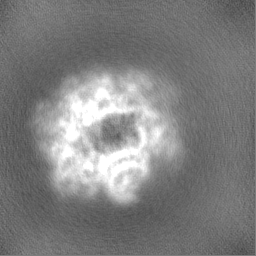
| Images |  emd_61464.png emd_61464.png | 20.7 KB | ||
| Filedesc metadata | emd-61464.cif.gz | 5.7 KB | ||
| Others | emd_61464_half_map_1.map.gzemd_61464_half_map_2.map.gz | 59.4 MB 59.4 MB | ||
| Archive directory |  http://ftp.pdbj.org/pub/emdb/structures/EMD-61464ftp://ftp.pdbj.org/pub/emdb/structures/EMD-61464 http://ftp.pdbj.org/pub/emdb/structures/EMD-61464ftp://ftp.pdbj.org/pub/emdb/structures/EMD-61464 | HTTPS FTP |
-Validation report
| Summary document | emd_61464_validation.pdf.gz | 949.4 KB | Display | EMDB validaton report |
|---|---|---|---|---|
| Full document | emd_61464_full_validation.pdf.gz | 949 KB | Display | |
| Data in XML | emd_61464_validation.xml.gz | 12.3 KB | Display | |
| Data in CIF | emd_61464_validation.cif.gz | 14.5 KB | Display | |
| Arichive directory | https://ftp.pdbj.org/pub/emdb/validation_reports/EMD-61464ftp://ftp.pdbj.org/pub/emdb/validation_reports/EMD-61464 | HTTPS FTP |
-Related structure data
| Related structure data |  9jghMC  9jgiC M: atomic model generated by this map C: citing same article ( |
|---|---|
| Similar structure data |

-Links
| EMDB pages | EMDB (EBI/PDBe) / EMDataResource |
|---|
-Map
| File | Download / File: emd_61464.map.gz / Format: CCP4 / Size: 64 MB / Type: IMAGE STORED AS FLOATING POINT NUMBER (4 BYTES) | ||||||||||||||||||||||||||||||||||||
|---|---|---|---|---|---|---|---|---|---|---|---|---|---|---|---|---|---|---|---|---|---|---|---|---|---|---|---|---|---|---|---|---|---|---|---|---|---|
| Annotation | cryo-EM structure of the TTP polymer tube's end | ||||||||||||||||||||||||||||||||||||
| Projections & slices | Image control
Images are generated by Spider. | ||||||||||||||||||||||||||||||||||||
| Voxel size | X=Y=Z: 0.83 Å | ||||||||||||||||||||||||||||||||||||
| Density |
| ||||||||||||||||||||||||||||||||||||
| Symmetry | Space group: 1 | ||||||||||||||||||||||||||||||||||||
| Details | EMDB XML:
|
 Z (Sec.)
 Y (Row.)
 X (Col.)


-Supplemental data
-Half map: cryo-EM structure of the TTP polymer tube's end
| File | emd_61464_half_map_1.map | ||||||||||||
|---|---|---|---|---|---|---|---|---|---|---|---|---|---|
| Annotation | cryo-EM structure of the TTP polymer tube's end | ||||||||||||
| Projections & Slices |
| ||||||||||||
| Density Histograms |


-Half map: cryo-EM structure of the TTP polymer tube's end
| File | emd_61464_half_map_2.map | ||||||||||||
|---|---|---|---|---|---|---|---|---|---|---|---|---|---|
| Annotation | cryo-EM structure of the TTP polymer tube's end | ||||||||||||
| Projections & Slices |
| ||||||||||||
| Density Histograms |


- Sample components
-Entire : cryo-EM structure of the TTP polymer at the tube's end
| Entire | Name: cryo-EM structure of the TTP polymer at the tube's end |
|---|---|
| Components |
|
-Supramolecule #1: cryo-EM structure of the TTP polymer at the tube's end
| Supramolecule | Name: cryo-EM structure of the TTP polymer at the tube's end type: complex / ID: 1 / Parent: 0 / Macromolecule list: all |
|---|---|
| Source (natural) | Organism: |
-Macromolecule #1: tube tail protein
| Macromolecule | Name: tube tail protein / type: protein_or_peptide / ID: 1 / Number of copies: 15 / Enantiomer: LEVO |
|---|---|
| Source (natural) | Organism: |
| Molecular weight | Theoretical: 30.133572 KDa |
| Recombinant expression | Organism: |
| Sequence | String: MKTVIQDTAD VYFKRKSDGK LVFTAEAQTA SFSQAISEEK LRGGIGNKPL YILKSEKEIN LTVKNAFFDL EWLAMTQGET IQEETKVKV FDREHGLIVD DTNKVTLKGK PVSDVTFYNK KGLTYKIAVS TDGTYTIPTA FAAAKDKLTA VYQIEKVGRR L AIKASKFS ...String: MKTVIQDTAD VYFKRKSDGK LVFTAEAQTA SFSQAISEEK LRGGIGNKPL YILKSEKEIN LTVKNAFFDL EWLAMTQGET IQEETKVKV FDREHGLIVD DTNKVTLKGK PVSDVTFYNK KGLTYKIAVS TDGTYTIPTA FAAAKDKLTA VYQIEKVGRR L AIKASKFS ERYEVEYRTI AYNPDTEEVY SDIYIQFPNV SPSGEFEMSL ENGNALAPEI KFEALADTDT DEMAVVIEAS RD ENTAAPV EDTTGSTQSS DLGGTTEHHH HHH UniProtKB: Uncharacterized protein |
-Experimental details
-Structure determination
| Method | negative staining, cryo EM |
|---|---|
 Processing Processing | single particle reconstruction |
| Aggregation state | filament |
-Sample preparation
| Buffer | pH: 8 Component:
Details: 40 mM Tris HCl, pH=8.0, 10 mM imidazole, 150 mM NaCl, and 1 mM phenylmethylsulfonyl fluoride PMSF | ||||||||||
|---|---|---|---|---|---|---|---|---|---|---|---|
| Staining | Type: NEGATIVE / Material: Uranyl Acetate | ||||||||||
| Vitrification | Cryogen name: ETHANE / Chamber humidity: 100 % / Chamber temperature: 4 K / Instrument: FEI VITROBOT MARK IV |
- Electron microscopy
| Microscope | FEI TECNAI SPIRIT |
|---|---|
| Image recording | Film or detector model: GATAN K3 (6k x 4k) / Average electron dose: 60.0 e/Å2 |
| Electron beam | Acceleration voltage: 300 kV / Electron source:  FIELD EMISSION GUN FIELD EMISSION GUN |
| Electron optics | Calibrated defocus max: 2.5 µm / Calibrated defocus min: 1.5 µm / Illumination mode: SPOT SCAN / Imaging mode: BRIGHT FIELD / Cs: 2.7 mm / Nominal defocus max: 2.5 µm / Nominal defocus min: 1.5 µm |
| Sample stage | Specimen holder model: FEI TITAN KRIOS AUTOGRID HOLDER / Cooling holder cryogen: NITROGEN |
| Experimental equipment |  Model: Tecnai Spirit / Image courtesy: FEI Company |
