 Movie
Movie Controller

[English] 日本語
 Yorodumi
- EMDB-6108: A molecular ruler determines the repeat length in eukaryotic cili... -
+ Open data

- Basic information
| Entry | Database: EMDB / ID: EMD-6108 | |||||||||
|---|---|---|---|---|---|---|---|---|---|---|
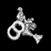
| Title | A molecular ruler determines the repeat length in eukaryotic cilia and flagella | |||||||||
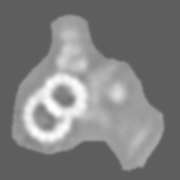
 Map data Map data | Wild-type axoneme | |||||||||
 Sample Sample |
| |||||||||
 Keywords Keywords | cryo-electron tomography / cilia and flagella / molecular ruler / FAP59 / FAP172 / CCDC39 / CCDC40 | |||||||||
| Biological species |   Chlamydomonas reinhardtii (plant) Chlamydomonas reinhardtii (plant) | |||||||||
| Method | subtomogram averaging / cryo EM / Resolution: 45.0 Å | |||||||||
 Authors Authors | Oda T / Yanagisawa H / Kamiya R / Kikkawa M | |||||||||
 Citation Citation | Journal: Science / Year: 2014 Title: A molecular ruler determines the repeat length in eukaryotic cilia and flagella. Authors: Toshiyuki Oda / Haruaki Yanagisawa / Ritsu Kamiya / Masahide Kikkawa /  Abstract: Existence of cellular structures with specific size raises a fundamental question in biology: How do cells measure length? One conceptual answer to this question is by a molecular ruler, but examples ...Existence of cellular structures with specific size raises a fundamental question in biology: How do cells measure length? One conceptual answer to this question is by a molecular ruler, but examples of such rulers in eukaryotes are lacking. In this work, we identified a molecular ruler in eukaryotic cilia and flagella. Using cryo-electron tomography, we found that FAP59 and FAP172 form a 96-nanometer (nm)-long complex in Chlamydomonas flagella and that the absence of the complex disrupted 96-nm repeats of axonemes. Furthermore, lengthening of the FAP59/172 complex by domain duplication resulted in extension of the repeats up to 128 nm, as well as duplication of specific axonemal components. Thus, the FAP59/172 complex is the molecular ruler that determines the 96-nm repeat length and arrangements of components in cilia and flagella. | |||||||||
| History |
|
- Structure visualization
| Movie |
 Movie viewer Movie viewer |
|---|---|
| Structure viewer | EM map: SurfViewMolmilJmol/JSmol |
| Supplemental images |

- Downloads & links
-EMDB archive
| Map data | emd_6108.map.gz | 43.2 MB | EMDB map data format | |
|---|---|---|---|---|
| Header (meta data) | emd-6108-v30.xmlemd-6108.xml | 8.4 KB 8.4 KB | Display Display | EMDB header |
| Images |  400_6108.gif 400_6108.gif 80_6108.gif 80_6108.gif | 39.1 KB 3.1 KB | ||
| Archive directory |  http://ftp.pdbj.org/pub/emdb/structures/EMD-6108ftp://ftp.pdbj.org/pub/emdb/structures/EMD-6108 http://ftp.pdbj.org/pub/emdb/structures/EMD-6108ftp://ftp.pdbj.org/pub/emdb/structures/EMD-6108 | HTTPS FTP |
-Related structure data
| Related structure data |  6109C 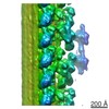 6110C 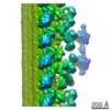 6111C 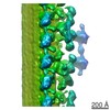 6112C 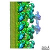 6113C 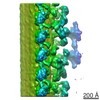 6114C  6115C  6116C 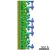 6117C C: citing same article ( |
|---|---|
| Similar structure data |
-Links
| EMDB pages | EMDB (EBI/PDBe) / EMDataResource |
|---|
-Map
| File | Download / File: emd_6108.map.gz / Format: CCP4 / Size: 48.3 MB / Type: IMAGE STORED AS FLOATING POINT NUMBER (4 BYTES) | ||||||||||||||||||||||||||||||||||||||||||||||||||||||||||||||||||||
|---|---|---|---|---|---|---|---|---|---|---|---|---|---|---|---|---|---|---|---|---|---|---|---|---|---|---|---|---|---|---|---|---|---|---|---|---|---|---|---|---|---|---|---|---|---|---|---|---|---|---|---|---|---|---|---|---|---|---|---|---|---|---|---|---|---|---|---|---|---|
| Annotation | Wild-type axoneme | ||||||||||||||||||||||||||||||||||||||||||||||||||||||||||||||||||||
| Projections & slices | Image control
Images are generated by Spider. generated in cubic-lattice coordinate | ||||||||||||||||||||||||||||||||||||||||||||||||||||||||||||||||||||
| Voxel size | X=Y=Z: 6.07 Å | ||||||||||||||||||||||||||||||||||||||||||||||||||||||||||||||||||||
| Density |
| ||||||||||||||||||||||||||||||||||||||||||||||||||||||||||||||||||||
| Symmetry | Space group: 1 | ||||||||||||||||||||||||||||||||||||||||||||||||||||||||||||||||||||
| Details | EMDB XML:
CCP4 map header:
| ||||||||||||||||||||||||||||||||||||||||||||||||||||||||||||||||||||
 Z (Sec.)
 Y (Row.)
 X (Col.)


-Supplemental data
- Sample components
-Entire : Wild-type axoneme from Chlamydomonas reinhardtii
| Entire | Name: Wild-type axoneme from Chlamydomonas reinhardtii |
|---|---|
| Components |
|
-Supramolecule #1000: Wild-type axoneme from Chlamydomonas reinhardtii
| Supramolecule | Name: Wild-type axoneme from Chlamydomonas reinhardtii / type: sample / ID: 1000 / Number unique components: 1 |
|---|
-Supramolecule #1: axoneme
| Supramolecule | Name: axoneme / type: organelle_or_cellular_component / ID: 1 / Recombinant expression: No / Database: NCBI |
|---|---|
| Source (natural) | Organism: Chlamydomonas reinhardtii (plant) / Strain: CC-125 / Organelle: Flagella |
-Experimental details
-Structure determination
| Method | cryo EM |
|---|---|
 Processing Processing | subtomogram averaging |
| Aggregation state | cell |
-Sample preparation
| Concentration | 0.01 mg/mL |
|---|---|
| Buffer | pH: 7.2 Details: 30 mM Hepes-NaOH, 5 mM MgCl2, 1 mM DTT, 1 mM EGTA, 50 mM NaCl |
| Grid | Details: 300 mesh copper grid with holey carbon support, glow discharged in the air |
| Vitrification | Cryogen name: ETHANE / Chamber humidity: 98 % / Chamber temperature: 93 K / Instrument: LEICA EM GP / Method: Blot for 5 seconds before plunging |
- Electron microscopy
| Microscope | JEOL 3100FFC |
|---|---|
| Specialist optics | Energy filter - Name: Omega / Energy filter - Lower energy threshold: 0.0 eV / Energy filter - Upper energy threshold: 20.0 eV |
| Date | Jun 1, 2014 |
| Image recording | Category: CCD / Film or detector model: TVIPS TEMCAM-F416 (4k x 4k) / Average electron dose: 100 e/Å2 |
| Electron beam | Acceleration voltage: 300 kV / Electron source:  FIELD EMISSION GUN FIELD EMISSION GUN |
| Electron optics | Illumination mode: FLOOD BEAM / Imaging mode: BRIGHT FIELD / Nominal defocus max: 7.0 µm / Nominal defocus min: 6.0 µm / Nominal magnification: 25700 |
| Sample stage | Specimen holder model: GATAN LIQUID NITROGEN / Tilt series - Axis1 - Min angle: -65 ° / Tilt series - Axis1 - Max angle: 65 ° |
-Image processing
| Details | Number of tilts (projections) used in 3D reconstruction: 65 Tomographic tilt angle increment: 2 |
|---|---|
| Final reconstruction | Resolution.type: BY AUTHOR / Resolution: 45.0 Å / Resolution method: OTHER |
