 Movie
Movie Controller


+ Open data

- Basic information
| Entry |  | |||||||||
|---|---|---|---|---|---|---|---|---|---|---|
| Title | cryo-EM structure of FtsE/X and ZipA complex in filament | |||||||||
 Map data Map data | ||||||||||
 Sample Sample |
| |||||||||
 Keywords Keywords | cell division / division filament / FtsE/X / ZipA / CELL CYCLE | |||||||||
| Function / homology |  Function and homology information Function and homology informationdivision septum / divisome complex / Gram-negative-bacterium-type cell wall / signal recognition particle binding / peptidoglycan turnover / SRP-dependent cotranslational protein targeting to membrane / plasma membrane protein complex / division septum assembly / FtsZ-dependent cytokinesis / cell division site ...division septum / divisome complex / Gram-negative-bacterium-type cell wall / signal recognition particle binding / peptidoglycan turnover / SRP-dependent cotranslational protein targeting to membrane / plasma membrane protein complex / division septum assembly / FtsZ-dependent cytokinesis / cell division site / ATPase complex / positive regulation of cell division / cell division / GTPase activity / GTP binding / protein homodimerization activity / ATP hydrolysis activity / ATP binding / membrane / plasma membrane Similarity search - Function | |||||||||
| Biological species |  | |||||||||
| Method | single particle reconstruction / cryo EM / Resolution: 3.3 Å | |||||||||
 Authors Authors | Zhu KF / Li JW / Luo M | |||||||||
| Funding support |  Singapore, 1 items Singapore, 1 items
| |||||||||
 Citation Citation | Journal: To Be Published Title: cryo-EM structure of FtsE/X and ZipA complex in filament Authors: Zhu KF / Li JW / Luo M | |||||||||
| History |
|
- Structure visualization
| Supplemental images |
|---|

- Downloads & links
-EMDB archive
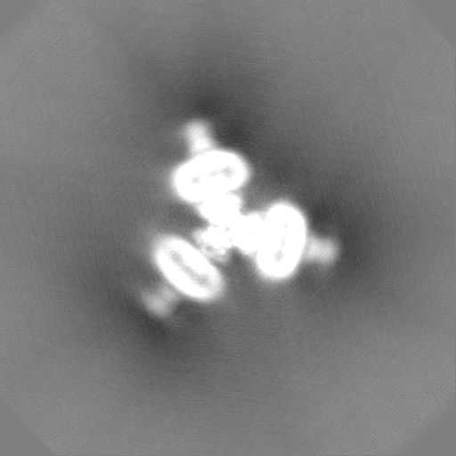
| Map data | emd_60898.map.gz | 483.7 MB | EMDB map data format | |
|---|---|---|---|---|
| Header (meta data) | emd-60898-v30.xmlemd-60898.xml | 20.3 KB 20.3 KB | Display Display | EMDB header |
| Images |  emd_60898.png emd_60898.png | 163.4 KB | ||
| Filedesc metadata | emd-60898.cif.gz | 6.3 KB | ||
| Others | emd_60898_half_map_1.map.gzemd_60898_half_map_2.map.gz | 462.2 MB 462.1 MB | ||
| Archive directory |  http://ftp.pdbj.org/pub/emdb/structures/EMD-60898ftp://ftp.pdbj.org/pub/emdb/structures/EMD-60898 http://ftp.pdbj.org/pub/emdb/structures/EMD-60898ftp://ftp.pdbj.org/pub/emdb/structures/EMD-60898 | HTTPS FTP |
-Related structure data
| Related structure data | 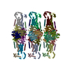 9iueMC M: atomic model generated by this map C: citing same article ( |
|---|---|
| Similar structure data |
-Links
| EMDB pages | EMDB (EBI/PDBe) / EMDataResource |
|---|---|
| Related items in Molecule of the Month |


-Map
| File | Download / File: emd_60898.map.gz / Format: CCP4 / Size: 512 MB / Type: IMAGE STORED AS FLOATING POINT NUMBER (4 BYTES) | ||||||||||||||||||||||||||||||||||||
|---|---|---|---|---|---|---|---|---|---|---|---|---|---|---|---|---|---|---|---|---|---|---|---|---|---|---|---|---|---|---|---|---|---|---|---|---|---|
| Projections & slices | Image control
Images are generated by Spider. | ||||||||||||||||||||||||||||||||||||
| Voxel size | X=Y=Z: 1.06 Å | ||||||||||||||||||||||||||||||||||||
| Density |
| ||||||||||||||||||||||||||||||||||||
| Symmetry | Space group: 1 | ||||||||||||||||||||||||||||||||||||
| Details | EMDB XML:
|
 Z (Sec.)
 Y (Row.)
 X (Col.)


-Supplemental data
-Half map: #2
| File | emd_60898_half_map_1.map | ||||||||||||
|---|---|---|---|---|---|---|---|---|---|---|---|---|---|
| Projections & Slices |
| ||||||||||||
| Density Histograms |


-Half map: #1
| File | emd_60898_half_map_2.map | ||||||||||||
|---|---|---|---|---|---|---|---|---|---|---|---|---|---|
| Projections & Slices |
| ||||||||||||
| Density Histograms |


- Sample components
-Entire : complex of FtsE/X-ZipA in filament
| Entire | Name: complex of FtsE/X-ZipA in filament |
|---|---|
| Components |
|
-Supramolecule #1: complex of FtsE/X-ZipA in filament
| Supramolecule | Name: complex of FtsE/X-ZipA in filament / type: complex / ID: 1 / Parent: 0 / Macromolecule list: #1-#3 |
|---|---|
| Source (natural) | Organism: |
-Macromolecule #1: Cell division protein ZipA
| Macromolecule | Name: Cell division protein ZipA / type: protein_or_peptide / ID: 1 / Number of copies: 18 / Enantiomer: LEVO |
|---|---|
| Source (natural) | Organism: |
| Molecular weight | Theoretical: 36.517199 KDa |
| Recombinant expression | Organism: |
| Sequence | String: MMQDLRLILI IVGAIAIIAL LVHGFWTSRK ERSSMFRDRP LKRMKSKRDD DSYDEDVEDD EGVGEVRVHR VNHAPANAQE HEAARPSPQ HQYQPPYASA QPRQPVQQPP EAQVPPQHAP HPAQPVQQPA YQPQPEQPLQ QPVSPQVAPA PQPVHSAPQP A QQAFQPAE ...String: MMQDLRLILI IVGAIAIIAL LVHGFWTSRK ERSSMFRDRP LKRMKSKRDD DSYDEDVEDD EGVGEVRVHR VNHAPANAQE HEAARPSPQ HQYQPPYASA QPRQPVQQPP EAQVPPQHAP HPAQPVQQPA YQPQPEQPLQ QPVSPQVAPA PQPVHSAPQP A QQAFQPAE PVAAPQPEPV AEPAPVMDKP KRKEAVIIMN VAAHHGSELN GELLLNSIQQ AGFIFGDMNI YHRHLSPDGS GP ALFSLAN MVKPGTFDPE MKDFTTPGVT IFMQVPSYGD ELQNFKLMLQ SAQHIADEVG GVVLDDQRRM MTPQKLREYQ DII REVKDA NA UniProtKB: Cell division protein ZipA |
-Macromolecule #2: Cell division ATP-binding protein FtsE
| Macromolecule | Name: Cell division ATP-binding protein FtsE / type: protein_or_peptide / ID: 2 / Number of copies: 18 / Enantiomer: LEVO |
|---|---|
| Source (natural) | Organism: |
| Molecular weight | Theoretical: 24.832695 KDa |
| Recombinant expression | Organism: |
| Sequence | String: SGLVMIRFEH VSKAYLGGRQ ALQGVTFHMQ PGEMAFLTGH SGAGKSTLLK LICGIERPSA GKIWFSGHDI TRLKNREVPF LRRQIGMIF QDHHLLMDRT VYDNVAIPLI IAGASGDDIR RRVSAALDKV GLLDKAKNFP IQLSGGEQQR VGIARAVVNK P AVLLADEP ...String: SGLVMIRFEH VSKAYLGGRQ ALQGVTFHMQ PGEMAFLTGH SGAGKSTLLK LICGIERPSA GKIWFSGHDI TRLKNREVPF LRRQIGMIF QDHHLLMDRT VYDNVAIPLI IAGASGDDIR RRVSAALDKV GLLDKAKNFP IQLSGGEQQR VGIARAVVNK P AVLLADEP TGNLDDALSE GILRLFEEFN RVGVTVLMAT HDINLISRRS YRMLTLSDGH LHGGVGHE UniProtKB: Cell division ATP-binding protein FtsE |
-Macromolecule #3: Cell division protein FtsX
| Macromolecule | Name: Cell division protein FtsX / type: protein_or_peptide / ID: 3 / Number of copies: 18 / Enantiomer: LEVO |
|---|---|
| Source (natural) | Organism: |
| Molecular weight | Theoretical: 38.5835 KDa |
| Recombinant expression | Organism: |
| Sequence | String: MNKRDAINHI RQFGGRLDRF RKSVGGSGDG GRNAPKRAKS SPKPVNRKTN VFNEQVRYAF HGALQDLKSK PFATFLTVMV IAISLTLPS VCYMVYKNVN QAATQYYPSP QITVYLQKTL DDDAAAGVVA QLQAEQGVEK VNYLSREDAL GEFRNWSGFG G ALDMLEEN ...String: MNKRDAINHI RQFGGRLDRF RKSVGGSGDG GRNAPKRAKS SPKPVNRKTN VFNEQVRYAF HGALQDLKSK PFATFLTVMV IAISLTLPS VCYMVYKNVN QAATQYYPSP QITVYLQKTL DDDAAAGVVA QLQAEQGVEK VNYLSREDAL GEFRNWSGFG G ALDMLEEN PLPAVAVVIP KLDFQGTESL NTLRDRITQI NGIDEVRMDD SWFARLAALT GLVGRVSAMI GVLMVAAVFL VI GNSVRLS IFARRDSINV QKLIGATDGF ILRPFLYGGA LLGFSGALLS LILSEILVLR LSSAVAEVAQ VFGTKFDING LSF DECLLL LLVCSMIGWV AAWLATVQHL RHFTPE UniProtKB: Cell division protein FtsX |
-Macromolecule #4: PHOSPHOTHIOPHOSPHORIC ACID-ADENYLATE ESTER
| Macromolecule | Name: PHOSPHOTHIOPHOSPHORIC ACID-ADENYLATE ESTER / type: ligand / ID: 4 / Number of copies: 18 / Formula: AGS |
|---|---|
| Molecular weight | Theoretical: 523.247 Da |
| Chemical component information |  ChemComp-AGS: |
-Experimental details
-Structure determination
| Method | cryo EM |
|---|---|
 Processing Processing | single particle reconstruction |
| Aggregation state | particle |
-Sample preparation
| Buffer | pH: 8 |
|---|---|
| Vitrification | Cryogen name: NITROGEN |
- Electron microscopy
| Microscope | TFS KRIOS |
|---|---|
| Image recording | #0 - Image recording ID: 1 / #0 - Film or detector model: GATAN K2 SUMMIT (4k x 4k) / #0 - Average electron dose: 40.0 e/Å2 / #1 - Image recording ID: 2 / #1 - Film or detector model: GATAN K2 SUMMIT (4k x 4k) / #1 - Average electron dose: 36.65 e/Å2 / #2 - Image recording ID: 3 / #2 - Film or detector model: GATAN K2 SUMMIT (4k x 4k) / #2 - Average electron dose: 36.65 e/Å2 |
| Electron beam | Acceleration voltage: 300 kV / Electron source:  FIELD EMISSION GUN FIELD EMISSION GUN |
| Electron optics | Illumination mode: FLOOD BEAM / Imaging mode: BRIGHT FIELD / Nominal defocus max: 2.5 µm / Nominal defocus min: 1.5 µm |
| Experimental equipment |  Model: Titan Krios / Image courtesy: FEI Company |

