Movie
Movie Controller

[English] 日本語
 Yorodumi
- EMDB-60696: Cucumber Green Mottle Mosaic Virus (CGMMV)coat protein assembly -
+ Open data

- Basic information
| Entry |  | |||||||||
|---|---|---|---|---|---|---|---|---|---|---|
| Title | Cucumber Green Mottle Mosaic Virus (CGMMV)coat protein assembly | |||||||||
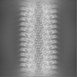
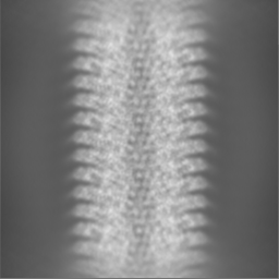
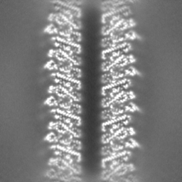
 Map data Map data | The map shows the helical arrangement of coat protein subunits of CGMMV. The RNA density, embedded inside the coat protein assembly, can be seen in higher contour level. | |||||||||
 Sample Sample |
| |||||||||
 Keywords Keywords | Helical / RNA virus / VIRUS | |||||||||
| Biological species |  Cucumber green mottle mosaic virus Cucumber green mottle mosaic virus | |||||||||
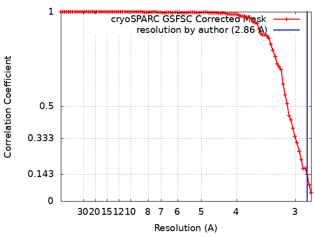
| Method | helical reconstruction / cryo EM / Resolution: 2.86 Å | |||||||||
 Authors Authors | Chatterjee A / Venkatasubramanian A / Jailani AK / Das U / Ragunath VK / Mandal B / Datta PP | |||||||||
| Funding support | 1 items
| |||||||||
 Citation Citation | Journal: To Be Published Title: Cucumber Green Mottle Mosaic Virus (CGMMV)coat protein assembly Authors: Chatterjee A / Venkatasubramanian A / Jailani AK / Das U / Ragunath VK / Mandal B / Datta PP | |||||||||
| History |
|
- Structure visualization
| Supplemental images |
|---|

- Downloads & links
-EMDB archive
| Map data | emd_60696.map.gz | 17 MB |  EMDB map data format EMDB map data format | |
|---|---|---|---|---|
| Header (meta data) | emd-60696-v30.xmlemd-60696.xml | 15.8 KB 15.8 KB | Display Display | EMDB header |
| FSC (resolution estimation) | emd_60696_fsc.xml | 8.4 KB | Display | FSC data file |
| Images |  emd_60696.png emd_60696.png | 66.1 KB | ||
| Masks | emd_60696_msk_1.map | 64 MB | Mask map | |
| Filedesc metadata | emd-60696.cif.gz | 4.8 KB | ||
| Others | emd_60696_half_map_1.map.gzemd_60696_half_map_2.map.gz | 57.7 MB 57.6 MB | ||
| Archive directory |  http://ftp.pdbj.org/pub/emdb/structures/EMD-60696ftp://ftp.pdbj.org/pub/emdb/structures/EMD-60696 http://ftp.pdbj.org/pub/emdb/structures/EMD-60696ftp://ftp.pdbj.org/pub/emdb/structures/EMD-60696 | HTTPS FTP |
-Related structure data
| Related structure data |  9vjd M: atomic model generated by this map |
|---|
-Links
| EMDB pages | EMDB (EBI/PDBe) / EMDataResource |
|---|
-Map
| File | Download / File: emd_60696.map.gz / Format: CCP4 / Size: 64 MB / Type: IMAGE STORED AS FLOATING POINT NUMBER (4 BYTES) | ||||||||||||||||||||||||||||||||||||
|---|---|---|---|---|---|---|---|---|---|---|---|---|---|---|---|---|---|---|---|---|---|---|---|---|---|---|---|---|---|---|---|---|---|---|---|---|---|
| Annotation | The map shows the helical arrangement of coat protein subunits of CGMMV. The RNA density, embedded inside the coat protein assembly, can be seen in higher contour level. | ||||||||||||||||||||||||||||||||||||
| Projections & slices | Image control
Images are generated by Spider. | ||||||||||||||||||||||||||||||||||||
| Voxel size | X=Y=Z: 1.38 Å | ||||||||||||||||||||||||||||||||||||
| Density |
| ||||||||||||||||||||||||||||||||||||
| Symmetry | Space group: 1 | ||||||||||||||||||||||||||||||||||||
| Details | EMDB XML:
|
 Z (Sec.)
 Y (Row.)
 X (Col.)


-Supplemental data
-Mask #1
| File | emd_60696_msk_1.map | ||||||||||||
|---|---|---|---|---|---|---|---|---|---|---|---|---|---|
| Projections & Slices |
| ||||||||||||
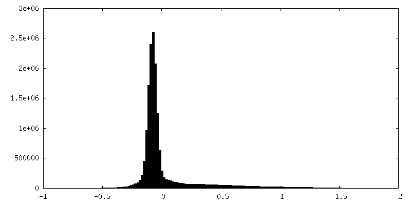
| Density Histograms |


-Half map: Half map B
| File | emd_60696_half_map_1.map | ||||||||||||
|---|---|---|---|---|---|---|---|---|---|---|---|---|---|
| Annotation | Half map B | ||||||||||||
| Projections & Slices |
| ||||||||||||
| Density Histograms |


-Half map: Half map A
| File | emd_60696_half_map_2.map | ||||||||||||
|---|---|---|---|---|---|---|---|---|---|---|---|---|---|
| Annotation | Half map A | ||||||||||||
| Projections & Slices |
| ||||||||||||
| Density Histograms |


- Sample components
-Entire : Cucumber green mottle mosaic virus
| Entire | Name: Cucumber green mottle mosaic virus |
|---|---|
| Components |
|
-Supramolecule #1: Cucumber green mottle mosaic virus
| Supramolecule | Name: Cucumber green mottle mosaic virus / type: virus / ID: 1 / Parent: 0 / Macromolecule list: all / NCBI-ID: 12235 / Sci species name: Cucumber green mottle mosaic virus / Sci species strain: Bg / Virus type: VIRION / Virus isolate: STRAIN / Virus enveloped: No / Virus empty: Yes |
|---|---|
| Host (natural) | Organism:  Lagenaria siceraria (white-flowered gourd) Lagenaria siceraria (white-flowered gourd) |
-Macromolecule #1: Cucumber Green Mottle Mosaic Virus
| Macromolecule | Name: Cucumber Green Mottle Mosaic Virus / type: other / ID: 1 / Classification: other |
|---|---|
| Source (natural) | Organism: Cucumber green mottle mosaic virus |
| Sequence | String: MAYNPITPSK LIAFSASYVP VRTLLNFLVA SQGTAFQTQA GRDSFRESLS ALPSSVVDIN SRFPDAGFYA FLNGPVLRPI FVSLLSSTDT RNRVIEVVDP SNPTTAESLN AVKRTDDAST AARAEIDNLI ESISKGFDVY DRASFEAAFS VVWSEATTSK A GENBANK: GENBANK: DQ767631.2 |
-Experimental details
-Structure determination
| Method | cryo EM |
|---|---|
 Processing Processing | helical reconstruction |
| Aggregation state | filament |
-Sample preparation
| Buffer | pH: 6.5 |
|---|---|
| Vitrification | Cryogen name: ETHANE |
- Electron microscopy
| Microscope | FEI TITAN KRIOS |
|---|---|
| Image recording | Film or detector model: FEI FALCON III (4k x 4k) / Average electron dose: 35.82 e/Å2 |
| Electron beam | Acceleration voltage: 300 kV / Electron source:  FIELD EMISSION GUN FIELD EMISSION GUN |
| Electron optics | Illumination mode: FLOOD BEAM / Imaging mode: BRIGHT FIELD / Nominal defocus max: 3.2 µm / Nominal defocus min: 1.8 µm / Nominal magnification: 59000 |
| Sample stage | Specimen holder model: FEI TITAN KRIOS AUTOGRID HOLDER / Cooling holder cryogen: NITROGEN |
| Experimental equipment |  Model: Titan Krios / Image courtesy: FEI Company |