[English] 日本語
 Yorodumi
- EMDB-60680: The Cryo-EM structure of MPXV E5 in complex with ssDNA in interme... -
+ Open data
- Basic information
| Entry |  | |||||||||
|---|---|---|---|---|---|---|---|---|---|---|
| Title | The Cryo-EM structure of MPXV E5 in complex with ssDNA in intermediate state 3 | |||||||||
 Map data Map data | ||||||||||
 Sample Sample |
| |||||||||
 Keywords Keywords | complex / VIRAL PROTEIN/DNA / VIRAL PROTEIN-DNA complex | |||||||||
| Function / homology |  Function and homology information Function and homology information | |||||||||
| Biological species |  Monkeypox virus Monkeypox virus | |||||||||
| Method | single particle reconstruction / cryo EM / Resolution: 3.12 Å | |||||||||
 Authors Authors | Cheng YX / Han P / Wang H | |||||||||
| Funding support |  China, 1 items China, 1 items
| |||||||||
 Citation Citation |  Journal: Nat Commun / Year: 2025 Journal: Nat Commun / Year: 2025Title: Assembly and breakage of head-to-head double hexamer reveals mpox virus E5-catalyzed DNA unwinding initiation. Authors: Yingxian Cheng / Pu Han / Qi Peng / Ruili Liu / Hao Liu / Bin Yuan / Yunfei Zhao / Lu Kuai / Jianxun Qi / Kai Miao / Yi Shi / George Fu Gao / Han Wang /  Abstract: The replicative helicase-catalyzed unwinding of the DNA double helix is the initiation of DNA replication. Helicases and primases are functionally related enzymes that have even been expressed as ...The replicative helicase-catalyzed unwinding of the DNA double helix is the initiation of DNA replication. Helicases and primases are functionally related enzymes that have even been expressed as fusion proteins in some organisms and viruses. However, the mechanism underlying DNA unwinding initiation by these helicase-primase fusion enzymes and the functional association between domains have not been elucidated. Herein, we report the cryo-EM structures of mpox virus E5, the founding member of these helicase-primase enzymes, in various enzymatic stages. Notably, E5 forms a head-to-head double hexamer encircling dsDNA, disrupted by the conformational rearrangement of primase domains upon nucleotide incorporation. Five E5-ssDNA-ATP structures further support an ATP cycle-driven non-classical escort model for E5 translocation. Finally, the helicase domain is found to enhance the primase function as a DNA scaffold. Together, our data shed light on the E5-mediated DNA unwinding model including dsDNA loading, DNA melting, ssDNA translocation, and provide a reasonable interpretation for evolutionary preservation of helicase-primase fusion from a functional perspective. | |||||||||
| History |
|
- Structure visualization
| Supplemental images |
|---|
- Downloads & links
-EMDB archive
| Map data |  emd_60680.map.gz emd_60680.map.gz | 210.5 MB |  EMDB map data format EMDB map data format | |
|---|---|---|---|---|
| Header (meta data) |  emd-60680-v30.xml emd-60680-v30.xml emd-60680.xml emd-60680.xml | 19 KB 19 KB | Display Display |  EMDB header EMDB header |
| FSC (resolution estimation) |  emd_60680_fsc.xml emd_60680_fsc.xml | 15.9 KB | Display |  FSC data file FSC data file |
| Images |  emd_60680.png emd_60680.png | 62 KB | ||
| Filedesc metadata |  emd-60680.cif.gz emd-60680.cif.gz | 6.6 KB | ||
| Others |  emd_60680_half_map_1.map.gz emd_60680_half_map_1.map.gz emd_60680_half_map_2.map.gz emd_60680_half_map_2.map.gz | 391.6 MB 391.6 MB | ||
| Archive directory |  http://ftp.pdbj.org/pub/emdb/structures/EMD-60680 http://ftp.pdbj.org/pub/emdb/structures/EMD-60680 ftp://ftp.pdbj.org/pub/emdb/structures/EMD-60680 ftp://ftp.pdbj.org/pub/emdb/structures/EMD-60680 | HTTPS FTP |
-Validation report
| Summary document |  emd_60680_validation.pdf.gz emd_60680_validation.pdf.gz | 885 KB | Display |  EMDB validaton report EMDB validaton report |
|---|---|---|---|---|
| Full document |  emd_60680_full_validation.pdf.gz emd_60680_full_validation.pdf.gz | 884.5 KB | Display | |
| Data in XML |  emd_60680_validation.xml.gz emd_60680_validation.xml.gz | 25.5 KB | Display | |
| Data in CIF |  emd_60680_validation.cif.gz emd_60680_validation.cif.gz | 33.2 KB | Display | |
| Arichive directory |  https://ftp.pdbj.org/pub/emdb/validation_reports/EMD-60680 https://ftp.pdbj.org/pub/emdb/validation_reports/EMD-60680 ftp://ftp.pdbj.org/pub/emdb/validation_reports/EMD-60680 ftp://ftp.pdbj.org/pub/emdb/validation_reports/EMD-60680 | HTTPS FTP |
-Related structure data
| Related structure data |  9im2MC  9ilyC  9ilzC  9im0C  9im1C  9im3C M: atomic model generated by this map C: citing same article ( |
|---|---|
| Similar structure data | Similarity search - Function & homology  F&H Search F&H Search |
- Links
Links
| EMDB pages |  EMDB (EBI/PDBe) / EMDB (EBI/PDBe) /  EMDataResource EMDataResource |
|---|---|
| Related items in Molecule of the Month |
- Map
Map
| File |  Download / File: emd_60680.map.gz / Format: CCP4 / Size: 421.9 MB / Type: IMAGE STORED AS FLOATING POINT NUMBER (4 BYTES) Download / File: emd_60680.map.gz / Format: CCP4 / Size: 421.9 MB / Type: IMAGE STORED AS FLOATING POINT NUMBER (4 BYTES) | ||||||||||||||||||||||||||||||||||||
|---|---|---|---|---|---|---|---|---|---|---|---|---|---|---|---|---|---|---|---|---|---|---|---|---|---|---|---|---|---|---|---|---|---|---|---|---|---|
| Projections & slices | Image control
Images are generated by Spider. | ||||||||||||||||||||||||||||||||||||
| Voxel size | X=Y=Z: 0.88 Å | ||||||||||||||||||||||||||||||||||||
| Density |
| ||||||||||||||||||||||||||||||||||||
| Symmetry | Space group: 1 | ||||||||||||||||||||||||||||||||||||
| Details | EMDB XML:
|
-Supplemental data
-Half map: #2
| File | emd_60680_half_map_1.map | ||||||||||||
|---|---|---|---|---|---|---|---|---|---|---|---|---|---|
| Projections & Slices |
| ||||||||||||
| Density Histograms |
-Half map: #1
| File | emd_60680_half_map_2.map | ||||||||||||
|---|---|---|---|---|---|---|---|---|---|---|---|---|---|
| Projections & Slices |
| ||||||||||||
| Density Histograms |
- Sample components
-Entire : MPXV E5 in complex with ssDNA in intermediate state 3
| Entire | Name: MPXV E5 in complex with ssDNA in intermediate state 3 |
|---|---|
| Components |
|
-Supramolecule #1: MPXV E5 in complex with ssDNA in intermediate state 3
| Supramolecule | Name: MPXV E5 in complex with ssDNA in intermediate state 3 / type: complex / ID: 1 / Parent: 0 / Macromolecule list: #1-#2 |
|---|---|
| Source (natural) | Organism:  Monkeypox virus Monkeypox virus |
-Macromolecule #1: Primase D5
| Macromolecule | Name: Primase D5 / type: protein_or_peptide / ID: 1 / Number of copies: 6 / Enantiomer: LEVO |
|---|---|
| Source (natural) | Organism:  Monkeypox virus Monkeypox virus |
| Molecular weight | Theoretical: 90.476344 KDa |
| Recombinant expression | Organism:  Spodoptera (butterflies/moths) Spodoptera (butterflies/moths) |
| Sequence | String: MDAAIRGNDV IFVLKTIGVP SACRQNEDPR FVEAFKCDEL ERYIDNNPEC TLFESLRDEE AYSIVRIFMD VDLDACLDEI DYLTAIQDF IIEVSNCVAR FAFTECGAIH ENVIKSMRSN FSLTKSTNRD KTSFHIIFLD TYTTMDTLIA MKRTLLELSR S SENPLTRS ...String: MDAAIRGNDV IFVLKTIGVP SACRQNEDPR FVEAFKCDEL ERYIDNNPEC TLFESLRDEE AYSIVRIFMD VDLDACLDEI DYLTAIQDF IIEVSNCVAR FAFTECGAIH ENVIKSMRSN FSLTKSTNRD KTSFHIIFLD TYTTMDTLIA MKRTLLELSR S SENPLTRS IDTAVYRRKT TLRVVGTRKN PNCDTIHVMQ PPHDNIEDYL FTYVDMNNNS YYFSLQRRLE DLVPDKLWEP GF ISFEDAI KRVSKIFINS IINFNDLDEN NFTTVPLVID YVTPCALCKK RSHKHPHQLS LENGAIRIYK TGNPHSCKVK IVP LDGNKL FNIAQRILDT NSVLLTERGD HIVWINNSWK FNSEEPLITK LILSIRHQLP KEYSSELLCP RKRKTVEANI RDML VDSVE TDTYPDKLPF KNGVLDLVDG MFYSGDDAKK YTCTVSTGFK FDDTKFVEDS PEMEELMNII NDIQPLTDEN KKNRE LYEK TLSSCLCGAT KGCLTFFFGE TATGKSTTKR LLKSAIGDLF VETGQTILTD VLDKGPNPFI ANMHLKRSVF CSELPD FAC SGSKKIRSDN IKKLTEPCVI GRPCFSNKIN NRNHATIIID TNYKPVFDRI DNALMRRIAV VRFRTHFSQP SGREAAE NN DAYDKVKLLD EGLDGKIQNN RYRFAFLYLL VKWYKKYHIP IMKLYPTPEE IPDFAFYLKI GTLLVSSSVK HIPLMTDL S KKGYILYDNV VTLPLTTFQQ KISKYFNSRL FGHDIESFIN RHKKFANVSD EYLQYIFIED ISSP UniProtKB: Uncoating factor OPG117 |
-Macromolecule #2: DNA (5'-D(P*TP*TP*TP*TP*TP*TP*T)-3')
| Macromolecule | Name: DNA (5'-D(P*TP*TP*TP*TP*TP*TP*T)-3') / type: dna / ID: 2 / Number of copies: 1 / Classification: DNA |
|---|---|
| Source (natural) | Organism:  Monkeypox virus Monkeypox virus |
| Molecular weight | Theoretical: 2.084392 KDa |
| Sequence | String: (DT)(DT)(DT)(DT)(DT)(DT)(DT) |
-Macromolecule #3: ADENOSINE MONOPHOSPHATE
| Macromolecule | Name: ADENOSINE MONOPHOSPHATE / type: ligand / ID: 3 / Number of copies: 1 / Formula: AMP |
|---|---|
| Molecular weight | Theoretical: 347.221 Da |
| Chemical component information |  ChemComp-AMP: |
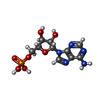
-Macromolecule #4: ADENOSINE-5'-TRIPHOSPHATE
| Macromolecule | Name: ADENOSINE-5'-TRIPHOSPHATE / type: ligand / ID: 4 / Number of copies: 3 / Formula: ATP |
|---|---|
| Molecular weight | Theoretical: 507.181 Da |
| Chemical component information |  ChemComp-ATP: |
-Macromolecule #5: MAGNESIUM ION
| Macromolecule | Name: MAGNESIUM ION / type: ligand / ID: 5 / Number of copies: 3 / Formula: MG |
|---|---|
| Molecular weight | Theoretical: 24.305 Da |
-Macromolecule #6: ADENOSINE-5'-DIPHOSPHATE
| Macromolecule | Name: ADENOSINE-5'-DIPHOSPHATE / type: ligand / ID: 6 / Number of copies: 1 / Formula: ADP |
|---|---|
| Molecular weight | Theoretical: 427.201 Da |
| Chemical component information |  ChemComp-ADP: |
-Experimental details
-Structure determination
| Method | cryo EM |
|---|---|
 Processing Processing | single particle reconstruction |
| Aggregation state | particle |
- Sample preparation
| Buffer | pH: 7.4 |
|---|---|
| Vitrification | Cryogen name: ETHANE |
- Electron microscopy
| Microscope | FEI TITAN KRIOS |
|---|---|
| Image recording | Film or detector model: GATAN K3 BIOQUANTUM (6k x 4k) / Average electron dose: 50.0 e/Å2 |
| Electron beam | Acceleration voltage: 300 kV / Electron source:  FIELD EMISSION GUN FIELD EMISSION GUN |
| Electron optics | Illumination mode: FLOOD BEAM / Imaging mode: BRIGHT FIELD / Nominal defocus max: 2.3000000000000003 µm / Nominal defocus min: 1.3 µm |
| Experimental equipment |  Model: Titan Krios / Image courtesy: FEI Company |
 Movie
Movie Controller


 Z (Sec.)
 Y (Row.)
 X (Col.)


