[English] 日本語
Yorodumi
- EMDB-60206: Structure of Polycystin-1/Polycystin-2 complex with phosphatidic ... -

+
Open data


ID or keywords:

Loading...

-
Basic information

Entry
Database: EMDB / ID: EMD-60206
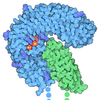
TitleStructure of Polycystin-1/Polycystin-2 complex with phosphatidic acid bound
Map data
Sample
  • Complex: Structure of Polycystin-1/Polycystin-2 complex with phosphatidic acid bound
    • Protein or peptide: Polycystin-1
    • Protein or peptide: Polycystin-2
  • Ligand: 2-acetamido-2-deoxy-beta-D-glucopyranose
  • Ligand: CALCIUM ION
  • Ligand: 1,2-DIOCTANOYL-SN-GLYCERO-3-PHOSPHATE
Keywordsion channel / MEMBRANE PROTEIN
Function / homology
Function and homology information


metanephric distal tubule morphogenesis / nitrogen cycle metabolic process / detection of nodal flow / metanephric smooth muscle tissue development / metanephric cortex development / metanephric cortical collecting duct development / metanephric distal tubule development / polycystin complex / mesonephric tubule development / mesonephric duct development ...metanephric distal tubule morphogenesis / nitrogen cycle metabolic process / detection of nodal flow / metanephric smooth muscle tissue development / metanephric cortex development / metanephric cortical collecting duct development / metanephric distal tubule development / polycystin complex / mesonephric tubule development / mesonephric duct development / metanephric part of ureteric bud development / renal tubule morphogenesis / determination of liver left/right asymmetry / HLH domain binding / lung epithelium development / metanephric ascending thin limb development / lymph vessel morphogenesis / metanephric mesenchyme development / metanephric proximal tubule development / metanephric S-shaped body morphogenesis / basal cortex / renal artery morphogenesis / mitocytosis / positive regulation of inositol 1,4,5-trisphosphate-sensitive calcium-release channel activity / calcium-induced calcium release activity / calcium-independent cell-matrix adhesion / Wnt receptor activity / migrasome / cilium organization / VxPx cargo-targeting to cilium / genitalia development / detection of mechanical stimulus / muscle alpha-actinin binding / regulation of calcium ion import / voltage-gated monoatomic ion channel activity / placenta blood vessel development / response to fluid shear stress / Golgi-associated vesicle membrane / cellular response to hydrostatic pressure / cation channel complex / cellular response to fluid shear stress / metanephric collecting duct development / outward rectifier potassium channel activity / actinin binding / cellular response to osmotic stress / non-motile cilium / digestive tract development / determination of left/right symmetry / inorganic cation transmembrane transport / voltage-gated monoatomic cation channel activity / aorta development / cartilage development / neural tube development / motile cilium / voltage-gated sodium channel activity / ciliary membrane / cartilage condensation / branching involved in ureteric bud morphogenesis / protein heterotetramerization / branching morphogenesis of an epithelial tube / skin development / negative regulation of G1/S transition of mitotic cell cycle / spinal cord development / establishment of cell polarity / transcription regulator inhibitor activity / cytoplasmic side of endoplasmic reticulum membrane / homophilic cell adhesion via plasma membrane adhesion molecules / heart looping / regulation of G1/S transition of mitotic cell cycle / centrosome duplication / voltage-gated potassium channel activity / cell surface receptor signaling pathway via JAK-STAT / anatomical structure morphogenesis / potassium channel activity / lateral plasma membrane / embryonic placenta development / voltage-gated calcium channel activity / regulation of cell adhesion / monoatomic cation channel activity / cytoskeletal protein binding / cellular response to cAMP / regulation of proteasomal protein catabolic process / release of sequestered calcium ion into cytosol / potassium ion transmembrane transport / calcium channel complex / sodium ion transmembrane transport / regulation of mitotic spindle organization / cytoplasmic vesicle membrane / cellular response to calcium ion / protein export from nucleus / liver development / basal plasma membrane / cell-matrix adhesion / kidney development / lumenal side of endoplasmic reticulum membrane / cellular response to reactive oxygen species / establishment of localization in cell / phosphoprotein binding / protein tetramerization / calcium ion transmembrane transport
Similarity search - Function
Polycystic kidney disease type 1 protein / PKD/REJ-like domain / Polycystin cation channel / REJ domain / REJ domain / REJ domain profile. / Polycystin-1 like, PLAT/LH2 domain / Carbohydrate-binding WSC / WSC domain / WSC domain profile. ...Polycystic kidney disease type 1 protein / PKD/REJ-like domain / Polycystin cation channel / REJ domain / REJ domain / REJ domain profile. / Polycystin-1 like, PLAT/LH2 domain / Carbohydrate-binding WSC / WSC domain / WSC domain profile. / present in yeast cell wall integrity and stress response component proteins / PKD domain / Ferredoxin I 4Fe-4S cluster domain / : / Polycystic kidney disease type 2 protein / Polycystin domain / Polycystin domain / Lipoxygenase homology 2 (beta barrel) domain / PLAT/LH2 domain / PLAT/LH2 domain superfamily / PLAT/LH2 domain / PLAT domain profile. / Polycystic kidney disease (PKD) domain profile. / Polycystin cation channel, PKD1/PKD2 / Polycystin cation channel / GPS motif / GAIN-B domain profile. / G-protein-coupled receptor proteolytic site domain / PKD domain / PKD domain superfamily / PKD/Chitinase domain / Repeats in polycystic kidney disease 1 (PKD1) and other proteins / Leucine-rich repeat N-terminal domain / Leucine rich repeat N-terminal domain / Cysteine-rich flanking region, C-terminal / Leucine rich repeat C-terminal domain / Lectin C-type domain / C-type lectin domain profile. / C-type lectin-like / C-type lectin (CTL) or carbohydrate-recognition domain (CRD) / C-type lectin-like/link domain superfamily / C-type lectin fold / Voltage-dependent channel domain superfamily / Leucine rich repeat / Leucine-rich repeat, typical subtype / Leucine-rich repeats, typical (most populated) subfamily / Leucine-rich repeat profile. / Leucine-rich repeat / Leucine-rich repeat domain superfamily / EF-hand calcium-binding domain profile. / EF-hand domain / EF-hand domain pair / Immunoglobulin-like fold
Similarity search - Domain/homology
Polycystin-1 / Polycystin-2
Similarity search - Component
Biological speciesHomo sapiens (human)
Methodsingle particle reconstruction / Resolution: 2.8 Å
AuthorsChen MY / Su Q / Shi YG / Yu Y
Funding support China, 2 items
OrganizationGrant numberCountry
National Natural Science Foundation of China (NSFC)31930059 China
National Natural Science Foundation of China (NSFC)81970633 China
CitationJournal: To Be Published
Title: Structure of Polycystin-1/Polycystin-2 complex with phosphatidic acid bound
Authors: Chen MY / Su Q / Shi YG
History
DepositionMay 17, 2024-
Header (metadata) releaseMay 21, 2025-
Map releaseMay 21, 2025-
UpdateMay 21, 2025-
Current statusMay 21, 2025Processing site: PDBc / Status: Released

-
Structure visualization

Supplemental images

Downloads & links

-
Map

FileDownload / File: emd_60206.map.gz / Format: CCP4 / Size: 52.7 MB / Type: IMAGE STORED AS FLOATING POINT NUMBER (4 BYTES)
Projections & slices

Image control

Size
Brightness
Contrast
Others
AxesZ (Sec.)Y (Row.)X (Col.)
1.09 Å/pix.
x 240 pix.
= 260.88 Å
1.09 Å/pix.
x 240 pix.
= 260.88 Å
1.09 Å/pix.
x 240 pix.
= 260.88 Å

Surface

Projections

Slices (1/3)

Slices (1/2)

Slices (2/3)

Images are generated by Spider.

Voxel sizeX=Y=Z: 1.087 Å
Density
Contour LevelBy AUTHOR: 0.45
Minimum - Maximum-2.774129 - 4.5372095
Average (Standard dev.)0.008850145 (±0.14021389)
SymmetrySpace group: 1
Details

EMDB XML:

Map geometry
Axis orderXYZ
Origin000
Dimensions240240240
Spacing240240240
CellA=B=C: 260.88 Å
α=β=γ: 90.0 °

-
Supplemental data

-
Half map: #1

Fileemd_60206_half_map_1.map
Projections & Slices
AxesZYX

Projections

Slices (1/2)
Density Histograms

-
Half map: #2

Fileemd_60206_half_map_2.map
Projections & Slices
AxesZYX

Projections

Slices (1/2)
Density Histograms

-
Sample components

-
Entire : Structure of Polycystin-1/Polycystin-2 complex with phosphatidic ...

EntireName: Structure of Polycystin-1/Polycystin-2 complex with phosphatidic acid bound
Components
  • Complex: Structure of Polycystin-1/Polycystin-2 complex with phosphatidic acid bound
    • Protein or peptide: Polycystin-1
    • Protein or peptide: Polycystin-2
  • Ligand: 2-acetamido-2-deoxy-beta-D-glucopyranose
  • Ligand: CALCIUM ION
  • Ligand: 1,2-DIOCTANOYL-SN-GLYCERO-3-PHOSPHATE

-
Supramolecule #1: Structure of Polycystin-1/Polycystin-2 complex with phosphatidic ...

SupramoleculeName: Structure of Polycystin-1/Polycystin-2 complex with phosphatidic acid bound
type: complex / ID: 1 / Parent: 0 / Macromolecule list: #1-#2
Source (natural)Organism: Homo sapiens (human)

-
Macromolecule #1: Polycystin-1

MacromoleculeName: Polycystin-1 / type: protein_or_peptide / ID: 1 / Number of copies: 1 / Enantiomer: LEVO
Source (natural)Organism: Homo sapiens (human)
Molecular weightTheoretical: 138.514672 KDa
Recombinant expressionOrganism: Homo sapiens (human)
SequenceString: MDYKDDDDKG ASLFVPPSHV RFVFPEPTAD VNYIVMLTCA VCLVTYMVMA AILHKLDQLD ASRGRAIPFC GQRGRFKYEI LVKTGWGRG SGTTAHVGIM LYGVDSRSGH RHLDGDRAFH RNSLDIFRIA TPHSLGSVWK IRVWHDNKGL SPAWFLQHVI V RDLQTARS ...String:
MDYKDDDDKG ASLFVPPSHV RFVFPEPTAD VNYIVMLTCA VCLVTYMVMA AILHKLDQLD ASRGRAIPFC GQRGRFKYEI LVKTGWGRG SGTTAHVGIM LYGVDSRSGH RHLDGDRAFH RNSLDIFRIA TPHSLGSVWK IRVWHDNKGL SPAWFLQHVI V RDLQTARS AFFLVNDWLS VETEANGGLV EKEVLAASDA ALLRFRRLLV AELQRGFFDK HIWLSIWDRP PRSRFTRIQR AT CCVLLIC LFLGANAVWY GAVGDSAYST GHVSRLSPLS VDTVAVGLVS SVVVYPVYLA ILFLFRMSRS KVAGSPSPTP AGQ QVLDID SCLDSSVLDS SFLTFSGLHA EQAFVGQMKS DLFLDDSKSL VCWPSGEGTL SWPDLLSDPS IVGSNLRQLA RGQA GHGLG PEEDGFSLAS PYSPAKSFSA SDEDLIQQVL AEGVSSPAPT QDTHMETDLL SSLSSTPGEK TETLALQRLG ELGPP SPGL NWEQPQAARL SRTGLVEGLR KRLLPAWCAS LAHGLSLLLV AVAVAVSGWV GASFPPGVSV AWLLSSSASF LASFLG WEP LKVLLEALYF SLVAKRLHPD EDDTLVESPA VTPVSARVPR VRPPHGFALF LAKEEARKVK RLHGMLRSLL VYMLFLL VT LLASYGDASC HGHAYRLQSA IKQELHSRAF LAITRSEELW PWMAHVLLPY VHGNQSSPEL GPPRLRQVRL QEALYPDP P GPRVHTCSAA GGFSTSDYDV GWESPHNGSG TWAYSAPDLL GAWSWGSCAV YDSGGYVQEL GLSLEESRDR LRFLQLHNW LDNRSRAVFL ELTRYSPAVG LHAAVTLRLE FPAAGRALAA LSVRPFALRR LSAGLSLPLL TSVCLLLFAV HFAVAEARTW HREGRWRVL RLGAWARWLL VALTAATALV RLAQLGAADR QWTRFVRGRP RRFTSFDQVA QLSSAARGLA ASLLFLLLVK A AQQLRFVR QWSVFGKTLC RALPELLGVT LGLVVLGVAY AQLAILLVSS CVDSLWSVAQ ALLVLCPGTG LSTLCPAESW HL SPLLCVG LWALRLWGAL RLGAVILRWR YHALRGELYR PAWEPQDYEM VELFLRRLRL WMGLSKVKEF RHKVRFEGME PLP SRSSRG SKVSPDVPPP SAGSDASHPS TSSSQLDGLS VSLGRLGTRC EPEPSRLQAV FEALLTQFDR LNQATEDVYQ LEQQ LHSLQ GRRSSRAPAG SSRGPSPGLR PALPSRLARA SRGVDLATGP SRTPLRAKNK VHPSST

UniProtKB: Polycystin-1

-
Macromolecule #2: Polycystin-2

MacromoleculeName: Polycystin-2 / type: protein_or_peptide / ID: 2 / Number of copies: 3 / Enantiomer: LEVO
Source (natural)Organism: Homo sapiens (human)
Molecular weightTheoretical: 113.555008 KDa
Recombinant expressionOrganism: Homo sapiens (human)
SequenceString: MGASSAWSHP QFEKGGGSGG GSGGSAWSHP QFEKGSAAAM VNSSRVQPQQ PGDAKRPPAP RAPDPGRLMA GCAAVGASLA APGGLCEQR GLEIEMQRIR QAAARDPPAG AAASPSPPLS SCSRQAWSRD NPGFEAEEEE EEVEGEEGGM VVEMDVEWRP G SRRSAASS ...String:
MGASSAWSHP QFEKGGGSGG GSGGSAWSHP QFEKGSAAAM VNSSRVQPQQ PGDAKRPPAP RAPDPGRLMA GCAAVGASLA APGGLCEQR GLEIEMQRIR QAAARDPPAG AAASPSPPLS SCSRQAWSRD NPGFEAEEEE EEVEGEEGGM VVEMDVEWRP G SRRSAASS AVSSVGARSR GLGGYHGAGH PSGRRRRRED QGPPCPSPVG GGDPLHRHLP LEGQPPRVAW AERLVRGLRG LW GTRLMEE SSTNREKYLK SVLRELVTYL LFLIVLCILT YGMMSSNVYY YTRMMSQLFL DTPVSKTEKT NFKTLSSMED FWK FTEGSL LDGLYWKMQP SNQTEADNRS FIFYENLLLG VPRIRQLRVR NGSCSIPQDL RDEIKECYDV YSVSSEDRAP FGPR NGTAW IYTSEKDLNG SSHWGIIATY SGAGYYLDLS RTREETAAQV ASLKKNVWLD RGTRATFIDF SVYNANINLF CVVRL LVEF PATGGVIPSW QFQPLKLIRY VTTFDFFLAA CEIIFCFFIF YYVVEEILEI RIHKLHYFRS FWNCLDVVIV VLSVVA IGI NIYRTSNVEV LLQFLEDQNT FPNFEHLAYW QIQFNNIAAV TVFFVWIKLF KFINFNRTMS QLSTTMSRCA KDLFGFA IM FFIIFLAYAQ LAYLVFGTQV DDFSTFQECI FTQFRIILGD INFAEIEEAN RVLGPIYFTT FVFFMFFILL NMFLAIIN D TYSEVKSDLA QQKAEMELSD LIRKGYHKAL VKLKLKKNTV DDISESLRQG GGKLNFDELR QDLKGKGHTD AEIEAIFTK YDQDGDQELT EHEHQQMRDD LEKEREDLDL DHSSLPRPMS SRSFPRSLDD SEEDDDEDSG HSSRRRGSIS SGVSYEEFQV LVRRVDRME HSIGSIVSKI DAVIVKLEIM ERAKLKRREV LGRLLDGVAE DERLGRDSEI HREQMERLVR EELERWESDD A ASQISHGL GTPVGLNGQP RPRSSRPSSS QSTEGMEGAG GNGSSNVHV

UniProtKB: Polycystin-2

-
Macromolecule #3: 2-acetamido-2-deoxy-beta-D-glucopyranose

MacromoleculeName: 2-acetamido-2-deoxy-beta-D-glucopyranose / type: ligand / ID: 3 / Number of copies: 8 / Formula: NAG
Molecular weightTheoretical: 221.208 Da
Chemical component information

ChemComp-NAG:
2-acetamido-2-deoxy-beta-D-glucopyranose

-
Macromolecule #4: CALCIUM ION

MacromoleculeName: CALCIUM ION / type: ligand / ID: 4 / Number of copies: 2 / Formula: CA
Molecular weightTheoretical: 40.078 Da

-
Macromolecule #5: 1,2-DIOCTANOYL-SN-GLYCERO-3-PHOSPHATE

MacromoleculeName: 1,2-DIOCTANOYL-SN-GLYCERO-3-PHOSPHATE / type: ligand / ID: 5 / Number of copies: 1 / Formula: PA8
Molecular weightTheoretical: 423.458 Da
Chemical component information

ChemComp-PA8:
1,2-DIOCTANOYL-SN-GLYCERO-3-PHOSPHATE

-
Experimental details

-
Structure determination

Processingsingle particle reconstruction
Aggregation stateparticle

-
Sample preparation

BufferpH: 7.4

-
Electron microscopy

MicroscopeFEI TITAN KRIOS
Image recordingFilm or detector model: GATAN K3 (6k x 4k) / Average electron dose: 50.0 e/Å2
Electron beamAcceleration voltage: 300 kV / Electron source: FIELD EMISSION GUN
Electron opticsIllumination mode: FLOOD BEAM / Imaging mode: BRIGHT FIELD / Nominal defocus max: 2.0 µm / Nominal defocus min: 1.2 µm
Experimental equipment
Model: Titan Krios / Image courtesy: FEI Company

+
Image processing

CTF correctionType: NONE
Startup modelType of model: PDB ENTRY
PDB model - PDB ID:
Final reconstructionResolution.type: BY AUTHOR / Resolution: 2.8 Å / Resolution method: FSC 0.143 CUT-OFF / Number images used: 146525
Initial angle assignmentType: MAXIMUM LIKELIHOOD
Final angle assignmentType: MAXIMUM LIKELIHOOD

+
About Yorodumi

-
News

-
Feb 9, 2022. New format data for meta-information of EMDB entries

New format data for meta-information of EMDB entries

  • Version 3 of the EMDB header file is now the official format.
  • The previous official version 1.9 will be removed from the archive.

Related info.:EMDB header

External links:wwPDB to switch to version 3 of the EMDB data model

-
Aug 12, 2020. Covid-19 info

Covid-19 info

URL: https://pdbj.org/emnavi/covid19.php

New page: Covid-19 featured information page in EM Navigator.

Related info.:Covid-19 info / Mar 5, 2020. Novel coronavirus structure data

+
Mar 5, 2020. Novel coronavirus structure data

Novel coronavirus structure data

Related info.:Yorodumi Speices / Aug 12, 2020. Covid-19 info

External links:COVID-19 featured content - PDBj / Molecule of the Month (242):Coronavirus Proteases

+
Jan 31, 2019. EMDB accession codes are about to change! (news from PDBe EMDB page)

EMDB accession codes are about to change! (news from PDBe EMDB page)

  • The allocation of 4 digits for EMDB accession codes will soon come to an end. Whilst these codes will remain in use, new EMDB accession codes will include an additional digit and will expand incrementally as the available range of codes is exhausted. The current 4-digit format prefixed with “EMD-” (i.e. EMD-XXXX) will advance to a 5-digit format (i.e. EMD-XXXXX), and so on. It is currently estimated that the 4-digit codes will be depleted around Spring 2019, at which point the 5-digit format will come into force.
  • The EM Navigator/Yorodumi systems omit the EMD- prefix.

Related info.:Q: What is EMD? / ID/Accession-code notation in Yorodumi/EM Navigator

External links:EMDB Accession Codes are Changing Soon! / Contact to PDBj

+
Jul 12, 2017. Major update of PDB

Major update of PDB

  • wwPDB released updated PDB data conforming to the new PDBx/mmCIF dictionary.
  • This is a major update changing the version number from 4 to 5, and with Remediation, in which all the entries are updated.
  • In this update, many items about electron microscopy experimental information are reorganized (e.g. em_software).
  • Now, EM Navigator and Yorodumi are based on the updated data.

External links:wwPDB Remediation / Enriched Model Files Conforming to OneDep Data Standards Now Available in the PDB FTP Archive

-
Yorodumi

Thousand views of thousand structures

  • Yorodumi is a browser for structure data from EMDB, PDB, SASBDB, etc.
  • This page is also the successor to EM Navigator detail page, and also detail information page/front-end page for Omokage search.
  • The word "yorodu" (or yorozu) is an old Japanese word meaning "ten thousand". "mi" (miru) is to see.

Related info.:EMDB / PDB / SASBDB / Comparison of 3 databanks / Yorodumi Search / Aug 31, 2016. New EM Navigator & Yorodumi / Yorodumi Papers / Jmol/JSmol / Function and homology information / Changes in new EM Navigator and Yorodumi

Read more