 Movie
Movie Controller

[English] 日本語
 Yorodumi
- EMDB-5989: Electron cryo-tomography of the Microtubule Nucleation Complex in... -
+ Open data

- Basic information
| Entry | Database: EMDB / ID: EMD-5989 | |||||||||
|---|---|---|---|---|---|---|---|---|---|---|
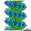
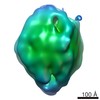
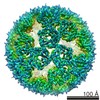
| Title | Electron cryo-tomography of the Microtubule Nucleation Complex in the Yeast Spindle Pole Body | |||||||||
 Map data Map data | Subtomogram average of the microtubule minus ends attached the purified yeast spindle pole body | |||||||||
 Sample Sample |
| |||||||||
 Keywords Keywords | microtubule / nucleation / gamma-tubulin / yeast | |||||||||
| Biological species |  | |||||||||
| Method | subtomogram averaging / cryo EM / Resolution: 38.0 Å | |||||||||
 Authors Authors | Kollman JM / Greenberg C / Li S / Fernandez JJ / Moritz M / Zelter A / Fong K / Sali A / Kilmartin JV / Davis TN / Agard DA | |||||||||
 Citation Citation | Journal: Nat Struct Mol Biol / Year: 2015 Title: Ring closure activates yeast γTuRC for species-specific microtubule nucleation. Authors: Justin M Kollman / Charles H Greenberg / Sam Li / Michelle Moritz / Alex Zelter / Kimberly K Fong / Jose-Jesus Fernandez / Andrej Sali / John Kilmartin / Trisha N Davis / David A Agard /    Abstract: The γ-tubulin ring complex (γTuRC) is the primary microtubule nucleator in cells. γTuRC is assembled from repeating γ-tubulin small complex (γTuSC) subunits and is thought to function as a ...The γ-tubulin ring complex (γTuRC) is the primary microtubule nucleator in cells. γTuRC is assembled from repeating γ-tubulin small complex (γTuSC) subunits and is thought to function as a template by presenting a γ-tubulin ring that mimics microtubule geometry. However, a previous yeast γTuRC structure showed γTuSC in an open conformation that prevents matching to microtubule symmetry. By contrast, we show here that γ-tubulin complexes are in a closed conformation when attached to microtubules. To confirm the functional importance of the closed γTuSC ring, we trapped the closed state and determined its structure, showing that the γ-tubulin ring precisely matches microtubule symmetry and providing detailed insight into γTuRC architecture. Importantly, the closed state is a stronger nucleator, thus suggesting that this conformational switch may allosterically control γTuRC activity. Finally, we demonstrate that γTuRCs have a strong preference for tubulin from the same species. | |||||||||
| History |
|
- Structure visualization
| Movie |
 Movie viewer Movie viewer |
|---|---|
| Structure viewer | EM map: SurfViewMolmilJmol/JSmol |
| Supplemental images |

- Downloads & links
-EMDB archive
| Map data | emd_5989.map.gz | 1.2 MB | EMDB map data format | |
|---|---|---|---|---|
| Header (meta data) | emd-5989-v30.xmlemd-5989.xml | 10.3 KB 10.3 KB | Display Display | EMDB header |
| Images |  400_5989.gif 400_5989.gif 80_5989.gif 80_5989.gif | 32.1 KB 2.8 KB | ||
| Archive directory |  http://ftp.pdbj.org/pub/emdb/structures/EMD-5989ftp://ftp.pdbj.org/pub/emdb/structures/EMD-5989 http://ftp.pdbj.org/pub/emdb/structures/EMD-5989ftp://ftp.pdbj.org/pub/emdb/structures/EMD-5989 | HTTPS FTP |
-Validation report
| Summary document | emd_5989_validation.pdf.gz | 79.3 KB | Display | EMDB validaton report |
|---|---|---|---|---|
| Full document | emd_5989_full_validation.pdf.gz | 78.4 KB | Display | |
| Data in XML | emd_5989_validation.xml.gz | 494 B | Display | |
| Arichive directory | https://ftp.pdbj.org/pub/emdb/validation_reports/EMD-5989ftp://ftp.pdbj.org/pub/emdb/validation_reports/EMD-5989 | HTTPS FTP |
-Related structure data

-Links
| EMDB pages | EMDB (EBI/PDBe) / EMDataResource |
|---|
-Map
| File | Download / File: emd_5989.map.gz / Format: CCP4 / Size: 1.9 MB / Type: IMAGE STORED AS FLOATING POINT NUMBER (4 BYTES) | ||||||||||||||||||||||||||||||||||||||||||||||||||||||||||||||||||||
|---|---|---|---|---|---|---|---|---|---|---|---|---|---|---|---|---|---|---|---|---|---|---|---|---|---|---|---|---|---|---|---|---|---|---|---|---|---|---|---|---|---|---|---|---|---|---|---|---|---|---|---|---|---|---|---|---|---|---|---|---|---|---|---|---|---|---|---|---|---|
| Annotation | Subtomogram average of the microtubule minus ends attached the purified yeast spindle pole body | ||||||||||||||||||||||||||||||||||||||||||||||||||||||||||||||||||||
| Projections & slices | Image control
Images are generated by Spider. | ||||||||||||||||||||||||||||||||||||||||||||||||||||||||||||||||||||
| Voxel size | X=Y=Z: 10.64 Å | ||||||||||||||||||||||||||||||||||||||||||||||||||||||||||||||||||||
| Density |
| ||||||||||||||||||||||||||||||||||||||||||||||||||||||||||||||||||||
| Symmetry | Space group: 1 | ||||||||||||||||||||||||||||||||||||||||||||||||||||||||||||||||||||
| Details | EMDB XML:
CCP4 map header:
| ||||||||||||||||||||||||||||||||||||||||||||||||||||||||||||||||||||
 Z (Sec.)
 Y (Row.)
 X (Col.)


-Supplemental data
- Sample components
-Entire : yeast spindle pole body
| Entire | Name: yeast spindle pole body |
|---|---|
| Components |
|
-Supramolecule #1000: yeast spindle pole body
| Supramolecule | Name: yeast spindle pole body / type: sample / ID: 1000 Oligomeric state: 7 gamma-TuSC complexes form a one-turn spiral Number unique components: 1 |
|---|
-Supramolecule #1: Spindle Pole Body
| Supramolecule | Name: Spindle Pole Body / type: organelle_or_cellular_component / ID: 1 / Name.synonym: microtubule organizing center / Recombinant expression: No / Database: NCBI |
|---|---|
| Source (natural) | Organism: |
-Experimental details
-Structure determination
| Method | cryo EM |
|---|---|
 Processing Processing | subtomogram averaging |
| Aggregation state | particle |
-Sample preparation
| Buffer | pH: 6.5 / Details: 10 mM Bis-Tris-Cl, 0.1 mM MgCl2, 20% v/v DMSO |
|---|---|
| Grid | Details: 300 mesh copper grid |
| Vitrification | Cryogen name: ETHANE / Chamber humidity: 90 % / Chamber temperature: 93 K / Instrument: FEI VITROBOT MARK III / Method: Blot for 3 seconds before plunging. |
- Electron microscopy
| Microscope | FEI POLARA 300 |
|---|---|
| Temperature | Average: 93 K |
| Alignment procedure | Legacy - Astigmatism: Objective lens astigmatism was corrected at 100,000 times magnification. |
| Specialist optics | Energy filter - Name: GIF / Energy filter - Lower energy threshold: 0.0 eV / Energy filter - Upper energy threshold: 25.0 eV |
| Date | May 14, 2010 |
| Image recording | Category: CCD / Film or detector model: OTHER / Digitization - Sampling interval: 1.5 µm / Number real images: 82 / Average electron dose: 60 e/Å2 / Bits/pixel: 8 |
| Electron beam | Acceleration voltage: 300 kV / Electron source:  FIELD EMISSION GUN FIELD EMISSION GUN |
| Electron optics | Calibrated magnification: 56391 / Illumination mode: FLOOD BEAM / Imaging mode: BRIGHT FIELD / Cs: 2.0 mm / Nominal defocus max: 1.5 µm / Nominal defocus min: 1.0 µm / Nominal magnification: 41000 |
| Sample stage | Specimen holder: Nitrogen-cooled / Specimen holder model: SIDE ENTRY, EUCENTRIC / Tilt series - Axis1 - Min angle: -60 ° / Tilt series - Axis1 - Max angle: 60 ° |
| Experimental equipment |  Model: Tecnai Polara / Image courtesy: FEI Company |
-Image processing
| Details | The subtomograms were selected manually. |
|---|---|
| Final reconstruction | Algorithm: OTHER / Resolution.type: BY AUTHOR / Resolution: 38.0 Å / Resolution method: OTHER / Software - Name: Xmipp / Number subtomograms used: 1156 |
| CTF correction | Details: each image, Wiener filter |
