 ムービー
ムービー コントローラー


+ データを開く

- 基本情報
基本情報
| 登録情報 | データベース: EMDB / ID: EMD-5950 | |||||||||
|---|---|---|---|---|---|---|---|---|---|---|
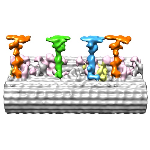
| タイトル | Cryo-electron tomography reveals ciliary defects underlying human RSPH1 primary ciliary dyskinesia | |||||||||
 マップデータ マップデータ | Reconstruction of 96 nm axonemal repeat of normal human respiratory cilia | |||||||||
 試料 試料 |
| |||||||||
 キーワード キーワード | axoneme / ciliopathy / human respiratory cilia / radial spoke / dynein | |||||||||
| 生物種 |  Homo sapiens (ヒト) Homo sapiens (ヒト) | |||||||||
| 手法 | サブトモグラム平均法 / クライオ電子顕微鏡法 / 解像度: 34.0 Å | |||||||||
 データ登録者 データ登録者 | Lin J / Yin W / Smith MC / Song KK / Leigh MW / Zariwala MA / Knowles MR / Ostrowski LE / Nicastro D | |||||||||
 引用 引用 | ジャーナル: Nat Commun / 年: 2014 タイトル: Cryo-electron tomography reveals ciliary defects underlying human RSPH1 primary ciliary dyskinesia. 著者: Jianfeng Lin / Weining Yin / Maria C Smith / Kangkang Song / Margaret W Leigh / Maimoona A Zariwala / Michael R Knowles / Lawrence E Ostrowski / Daniela Nicastro /  要旨: Cilia play essential roles in normal human development and health; cilia dysfunction results in diseases such as primary ciliary dyskinesia (PCD). Despite their importance, the native structure of ...Cilia play essential roles in normal human development and health; cilia dysfunction results in diseases such as primary ciliary dyskinesia (PCD). Despite their importance, the native structure of human cilia is unknown, and structural defects in the cilia of patients are often undetectable or remain elusive because of heterogeneity. Here we develop an approach that enables visualization of human (patient) cilia at high-resolution using cryo-electron tomography of samples obtained noninvasively by nasal scrape biopsy. We present the native 3D structures of normal and PCD-causing RSPH1-mutant human respiratory cilia in unprecedented detail; this allows comparisons of cilia structure across evolutionarily distant species and reveals the previously unknown primary defect and the heterogeneous secondary defects in RSPH1-mutant cilia. Our data provide evidence for structural and functional heterogeneity in radial spokes, suggest a mechanism for the milder RSPH1 PCD phenotype and demonstrate that cryo-electron tomography can be applied to human disease by directly imaging patient samples. | |||||||||
| 履歴 |
|
- 構造の表示
構造の表示
| ムービー |
 ムービービューア ムービービューア |
|---|---|
| 構造ビューア | EMマップ: SurfViewMolmilJmol/JSmol |
| 添付画像 |
- ダウンロードとリンク
-EMDBアーカイブ
| マップデータ | emd_5950.map.gz | 3.1 MB | EMDBマップデータ形式 | |
|---|---|---|---|---|
| ヘッダ (付随情報) | emd-5950-v30.xmlemd-5950.xml | 9.9 KB 9.9 KB | 表示 表示 | EMDBヘッダ |
| 画像 | emd_5950.tif | 173.3 KB | ||
| アーカイブディレクトリ |  http://ftp.pdbj.org/pub/emdb/structures/EMD-5950ftp://ftp.pdbj.org/pub/emdb/structures/EMD-5950 http://ftp.pdbj.org/pub/emdb/structures/EMD-5950ftp://ftp.pdbj.org/pub/emdb/structures/EMD-5950 | HTTPS FTP |
-検証レポート
| 文書・要旨 | emd_5950_validation.pdf.gz | 79.3 KB | 表示 | EMDB検証レポート |
|---|---|---|---|---|
| 文書・詳細版 | emd_5950_full_validation.pdf.gz | 78.4 KB | 表示 | |
| XML形式データ | emd_5950_validation.xml.gz | 494 B | 表示 | |
| アーカイブディレクトリ | https://ftp.pdbj.org/pub/emdb/validation_reports/EMD-5950ftp://ftp.pdbj.org/pub/emdb/validation_reports/EMD-5950 | HTTPS FTP |
-関連構造データ
| 類似構造データ |
|---|
-リンク
| EMDBのページ | EMDB (EBI/PDBe) / EMDataResource |
|---|
-マップ
| ファイル | ダウンロード / ファイル: emd_5950.map.gz / 形式: CCP4 / 大きさ: 3.9 MB / タイプ: IMAGE STORED AS FLOATING POINT NUMBER (4 BYTES) | ||||||||||||||||||||||||||||||||||||||||||||||||||||||||||||||||||||
|---|---|---|---|---|---|---|---|---|---|---|---|---|---|---|---|---|---|---|---|---|---|---|---|---|---|---|---|---|---|---|---|---|---|---|---|---|---|---|---|---|---|---|---|---|---|---|---|---|---|---|---|---|---|---|---|---|---|---|---|---|---|---|---|---|---|---|---|---|---|
| 注釈 | Reconstruction of 96 nm axonemal repeat of normal human respiratory cilia | ||||||||||||||||||||||||||||||||||||||||||||||||||||||||||||||||||||
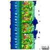
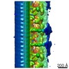
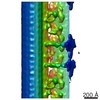
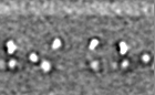
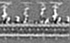
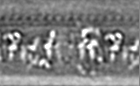
| 投影像・断面図 | 画像のコントロール
画像は Spider により作成 これらの図は立方格子座標系で作成されたものです | ||||||||||||||||||||||||||||||||||||||||||||||||||||||||||||||||||||
| ボクセルのサイズ | X=Y=Z: 9.997 Å | ||||||||||||||||||||||||||||||||||||||||||||||||||||||||||||||||||||
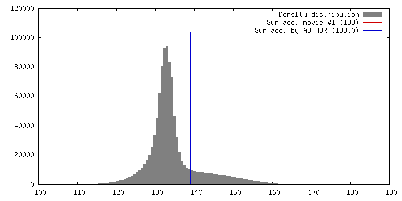
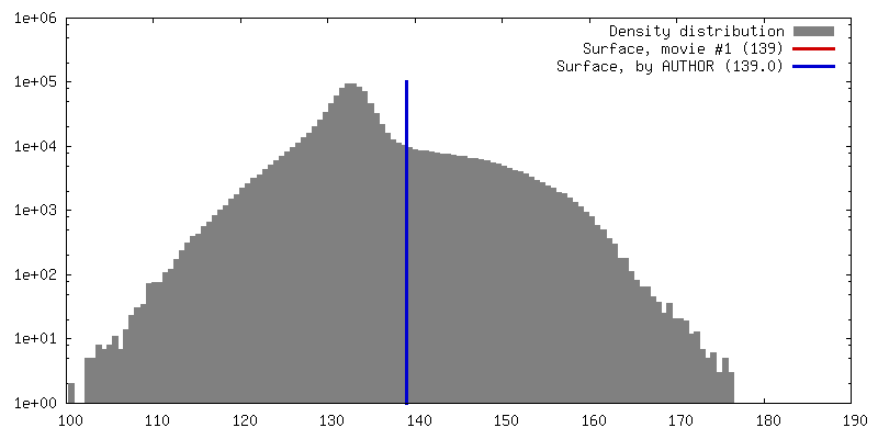
| 密度 |
| ||||||||||||||||||||||||||||||||||||||||||||||||||||||||||||||||||||
| 対称性 | 空間群: 1 | ||||||||||||||||||||||||||||||||||||||||||||||||||||||||||||||||||||
| 詳細 | EMDB XML:
CCP4マップ ヘッダ情報:
| ||||||||||||||||||||||||||||||||||||||||||||||||||||||||||||||||||||
 Z (Sec.)
 Y (Row.)
 X (Col.)


-添付データ
- 試料の構成要素
-全体 : Normal human respiratory ciliary axonemes
| 全体 | 名称: Normal human respiratory ciliary axonemes |
|---|---|
| 要素 |
|
-超分子 #1000: Normal human respiratory ciliary axonemes
| 超分子 | 名称: Normal human respiratory ciliary axonemes / タイプ: sample / ID: 1000 詳細: The samples were normal human respiratory ciliary axonemes isolated from cultures of trachea-bronchial epithelial cells. Number unique components: 1 |
|---|
-超分子 #1: Axoneme
| 超分子 | 名称: Axoneme / タイプ: organelle_or_cellular_component / ID: 1 / 組換発現: No / データベース: NCBI |
|---|---|
| 由来(天然) | 生物種: Homo sapiens (ヒト) / 別称: Human / 組織: Trachea-bronchial / 細胞: Epithelial cells / Organelle: Cilia |
-実験情報
-構造解析
| 手法 | クライオ電子顕微鏡法 |
|---|---|
 解析 解析 | サブトモグラム平均法 |
| 試料の集合状態 | cell |
-試料調製
| 緩衝液 | pH: 7.3 詳細: 30 mM Hepes, pH 7.3, 1 mM EGTA, 5 mM MgSO4, 0.1 mM EDTA, 25 mM NaCl, 1 mM dithiothreitol, 1% protease inhibitor cocktail, 100 g/mL soybean trypsin inhibitor (Sigma T9128) |
|---|---|
| グリッド | 詳細: Quantifoil holey carbon grids Cu 200 mesh R2/2 |
| 凍結 | 凍結剤: ETHANE / チャンバー内温度: 100 K / 装置: HOMEMADE PLUNGER / 手法: Blot for 1.5-2.5 seconds before plunging |
- 電子顕微鏡法
| 顕微鏡 | FEI TECNAI F30 |
|---|---|
| 特殊光学系 | エネルギーフィルター - 名称: GATAN postcolumn filter GIF エネルギーフィルター - エネルギー下限: 0.0 eV エネルギーフィルター - エネルギー上限: 20.0 eV |
| 日付 | 2013年2月5日 |
| 撮影 | カテゴリ: CCD フィルム・検出器のモデル: GATAN ULTRASCAN 1000 (2k x 2k) 平均電子線量: 100 e/Å2 |
| 電子線 | 加速電圧: 300 kV / 電子線源:  FIELD EMISSION GUN FIELD EMISSION GUN |
| 電子光学系 | 照射モード: FLOOD BEAM / 撮影モード: BRIGHT FIELD / 最大 デフォーカス(公称値): 8.0 µm / 最小 デフォーカス(公称値): 6.0 µm / 倍率(公称値): 13500 |
| 試料ステージ | 試料ホルダーモデル: GATAN LIQUID NITROGEN / Tilt series - Axis1 - Min angle: -65 ° / Tilt series - Axis1 - Max angle: 65 ° |
| 実験機器 |  モデル: Tecnai F30 / 画像提供: FEI Company |
-画像解析
| 詳細 | 3D tomograms were reconstructed using fiducial alignment and weighted backprojection using IMOD software. Final maps were calculated by averaging 950 particles (96-nm-long axonemal repeats) from 12 tomograms using the PEET software. |
|---|---|
| 最終 再構成 | アルゴリズム: OTHER / 解像度のタイプ: BY AUTHOR / 解像度: 34.0 Å / 解像度の算出法: OTHER / ソフトウェア - 名称: IMOD 詳細: Final map was calculated by averaging 850 particles (96-nm-long axonemal repeats) from 12 tomograms. 使用したサブトモグラム数: 950 |
