 Movie
Movie Controller

[English] 日本語
 Yorodumi
- EMDB-59306: EM map of in-cell structure of the membrane-bound chloroplast rib... -
+ Open data

- Basic information
| Entry |  | |||||||||
|---|---|---|---|---|---|---|---|---|---|---|
| Title | EM map of in-cell structure of the membrane-bound chloroplast ribosome in Chlamydomonas reinhardtii | |||||||||
 Map data Map data | ||||||||||
 Sample Sample |
| |||||||||
 Keywords Keywords | in-cell / chloroplast / ribosome / Chlamydomonas reinhardtii | |||||||||
| Biological species |  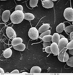 Chlamydomonas reinhardtii (plant) Chlamydomonas reinhardtii (plant) | |||||||||
| Method | subtomogram averaging / cryo EM / Resolution: 6.9 Å | |||||||||
 Authors Authors | Hou Z / Zhang P | |||||||||
| Funding support |  United Kingdom, 1 items United Kingdom, 1 items
| |||||||||
 Citation Citation | Journal: To Be Published Title: In-cell structural analysis reveals a distinctive chloroplast ribosome in Chlamydomonas reinhardtii Authors: Hou Z / Shen Y / Zhang Z / Lu P / Katzourakis A / Zhang P | |||||||||
| History |
|
- Structure visualization
| Supplemental images |
|---|

- Downloads & links
-EMDB archive
| Map data | emd_59306.map.gz | 8.5 MB |  EMDB map data format EMDB map data format | |
|---|---|---|---|---|
| Header (meta data) | emd-59306-v30.xmlemd-59306.xml | 16.3 KB 16.3 KB | Display Display | EMDB header |
| FSC (resolution estimation) | emd_59306_fsc.xml | 9.2 KB | Display | FSC data file |
| Images |  emd_59306.png emd_59306.png | 62.4 KB | ||
| Masks | emd_59306_msk_1.map | 64 MB | Mask map | |
| Filedesc metadata | emd-59306.cif.gz | 4.2 KB | ||
| Others | emd_59306_half_map_1.map.gzemd_59306_half_map_2.map.gz | 31.6 MB 31.6 MB | ||
| Archive directory |  https://data.pdbj.org/pub/emdb/structures/EMD-59306ftp://data.pdbj.org/pub/emdb/structures/EMD-59306 https://data.pdbj.org/pub/emdb/structures/EMD-59306ftp://data.pdbj.org/pub/emdb/structures/EMD-59306 | HTTPS FTP |
-Related structure data


-Links
| EMDB pages | EMDB (EBI/PDBe) / EMDataResource |
|---|
-Map
| File | Download / File: emd_59306.map.gz / Format: CCP4 / Size: 64 MB / Type: IMAGE STORED AS FLOATING POINT NUMBER (4 BYTES) | ||||||||||||||||||||||||||||||||||||
|---|---|---|---|---|---|---|---|---|---|---|---|---|---|---|---|---|---|---|---|---|---|---|---|---|---|---|---|---|---|---|---|---|---|---|---|---|---|
| Projections & slices | Image control
Images are generated by Spider. | ||||||||||||||||||||||||||||||||||||
| Voxel size | X=Y=Z: 1.903 Å | ||||||||||||||||||||||||||||||||||||
| Density |
| ||||||||||||||||||||||||||||||||||||
| Symmetry | Space group: 1 | ||||||||||||||||||||||||||||||||||||
| Details | EMDB XML:
|
 Z (Sec.)
 Y (Row.)
 X (Col.)


-Supplemental data
-Mask #1
| File | emd_59306_msk_1.map | ||||||||||||
|---|---|---|---|---|---|---|---|---|---|---|---|---|---|
| Projections & Slices |
| ||||||||||||
| Density Histograms |


-Half map: #1
| File | emd_59306_half_map_1.map | ||||||||||||
|---|---|---|---|---|---|---|---|---|---|---|---|---|---|
| Projections & Slices |
| ||||||||||||
| Density Histograms |


-Half map: #2
| File | emd_59306_half_map_2.map | ||||||||||||
|---|---|---|---|---|---|---|---|---|---|---|---|---|---|
| Projections & Slices |
| ||||||||||||
| Density Histograms |


- Sample components
-Entire : Wild-type Chlamydomonas reinhardtii cell
| Entire | Name: Wild-type Chlamydomonas reinhardtii cell |
|---|---|
| Components |
|
-Supramolecule #1: Wild-type Chlamydomonas reinhardtii cell
| Supramolecule | Name: Wild-type Chlamydomonas reinhardtii cell / type: cell / ID: 1 / Parent: 0 |
|---|---|
| Source (natural) | Organism: Chlamydomonas reinhardtii (plant) |
-Experimental details
-Structure determination
| Method | cryo EM |
|---|---|
 Processing Processing | subtomogram averaging |
| Aggregation state | cell |
-Sample preparation
| Buffer | pH: 7 |
|---|---|
| Grid | Model: Quantifoil R2/1 / Material: COPPER / Mesh: 300 |
| Vitrification | Cryogen name: ETHANE / Chamber humidity: 70 % / Chamber temperature: 298.15 K / Instrument: OTHER Details: The value given for _em_vitrification.instrument is LEICA EM GP2. This is not in a list of allowed values {'SPT LABTECH CHAMELEON', 'FEI VITROBOT MARK II', 'LEICA KF80', 'EMS-002 RAPID ...Details: The value given for _em_vitrification.instrument is LEICA EM GP2. This is not in a list of allowed values {'SPT LABTECH CHAMELEON', 'FEI VITROBOT MARK II', 'LEICA KF80', 'EMS-002 RAPID IMMERSION FREEZER', 'LEICA EM CPC', 'GATAN CRYOPLUNGE 3', 'ZEISS PLUNGE FREEZER CRYOBOX', 'FEI VITROBOT MARK IV', 'CRYOSOL VITROJET', 'FEI VITROBOT MARK III', 'FEI VITROBOT MARK I', 'HOMEMADE PLUNGER', 'LEICA PLUNGER', 'LEICA EM GP', 'REICHERT-JUNG PLUNGER', 'SPOTITON', 'OTHER'} so OTHER is written into the XML file. |
- Electron microscopy
| Microscope | TFS KRIOS |
|---|---|
| Specialist optics | Energy filter - Name: TFS Selectris / Energy filter - Slit width: 10 eV |
| Image recording | Film or detector model: TFS FALCON 4i (4k x 4k) / Average electron dose: 2.5 e/Å2 |
| Electron beam | Acceleration voltage: 300 kV / Electron source:  FIELD EMISSION GUN FIELD EMISSION GUN |
| Electron optics | C2 aperture diameter: 100.0 µm / Illumination mode: FLOOD BEAM / Imaging mode: BRIGHT FIELD / Cs: 2.7 mm / Nominal defocus max: 5.0 µm / Nominal defocus min: 2.0 µm / Nominal magnification: 64000 |
| Experimental equipment |  Model: Titan Krios / Image courtesy: FEI Company |

