[English] 日本語
Yorodumi
- EMDB-58849: R0-state of wild type human mitochondrial LONP1 protease bound to... -

+
Open data


ID or keywords:

Loading...

-
Basic information

Entry
Database: EMDB / ID: EMD-58849
TitleR0-state of wild type human mitochondrial LONP1 protease bound to endogenous ADP
Map data
Sample
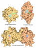
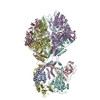
  • Complex: Lon protease homolog, mitochondrial
    • Protein or peptide: Lon protease homolog, mitochondrial
  • Ligand: ADENOSINE-5'-DIPHOSPHATE
KeywordsAAA+ protease / Lon protease / MOTOR PROTEIN
Function / homology
Function and homology information


oxidation-dependent protein catabolic process / response to aluminum ion / PH domain binding / endopeptidase La / mitochondrial protein catabolic process / G-quadruplex DNA binding / ATP-dependent peptidase activity / protein quality control for misfolded or incompletely synthesized proteins / mitochondrial nucleoid / insulin receptor substrate binding ...oxidation-dependent protein catabolic process / response to aluminum ion / PH domain binding / endopeptidase La / mitochondrial protein catabolic process / G-quadruplex DNA binding / ATP-dependent peptidase activity / protein quality control for misfolded or incompletely synthesized proteins / mitochondrial nucleoid / insulin receptor substrate binding / Mitochondrial unfolded protein response (UPRmt) / chaperone-mediated protein complex assembly / DNA polymerase binding / response to hormone / negative regulation of insulin receptor signaling pathway / Mitochondrial protein degradation / : / mitochondrion organization / ADP binding / single-stranded DNA binding / cellular response to oxidative stress / sequence-specific DNA binding / response to hypoxia / single-stranded RNA binding / mitochondrial matrix / serine-type endopeptidase activity / ATP hydrolysis activity / mitochondrion / nucleoplasm / ATP binding / membrane / identical protein binding / cytosol
Similarity search - Function
Lon protease homologue, chloroplastic/mitochondrial / : / Lon protease, bacterial/eukaryotic-type / Lon protease AAA+ ATPase lid domain / Peptidase S16, active site / ATP-dependent serine proteases, lon family, serine active site. / Lon proteolytic domain profile. / Peptidase S16, Lon proteolytic domain / Lon protease / Lon protease (S16) C-terminal proteolytic domain ...Lon protease homologue, chloroplastic/mitochondrial / : / Lon protease, bacterial/eukaryotic-type / Lon protease AAA+ ATPase lid domain / Peptidase S16, active site / ATP-dependent serine proteases, lon family, serine active site. / Lon proteolytic domain profile. / Peptidase S16, Lon proteolytic domain / Lon protease / Lon protease (S16) C-terminal proteolytic domain / Lon N-terminal domain profile. / Lon protease, N-terminal domain / Lon protease, N-terminal domain superfamily / ATP-dependent protease La (LON) substrate-binding domain / Found in ATP-dependent protease La (LON) / PUA-like superfamily / ATPase family associated with various cellular activities (AAA) / ATPase, AAA-type, core / Ribosomal protein S5 domain 2-type fold, subgroup / Ribosomal protein S5 domain 2-type fold / ATPases associated with a variety of cellular activities / AAA+ ATPase domain / P-loop containing nucleoside triphosphate hydrolase
Similarity search - Domain/homology
Lon protease homolog, mitochondrial
Similarity search - Component
Biological speciesHomo sapiens (human)
Methodsingle particle reconstruction / cryo EM / Resolution: 3.81 Å
AuthorsSchenck N / Roesgaard MA / Abrahams JP
Funding support Switzerland, 1 items
OrganizationGrant numberCountry
Other government Switzerland
CitationJournal: To Be Published
Title: Mitochondrial Lon protease couples substrate translocation to proteolytic activation
Authors: Schenck N / Roesgaard MA / Abrahams JP
History
DepositionJul 8, 2026-
Header (metadata) releaseJul 15, 2026-
Map releaseJul 15, 2026-
UpdateJul 15, 2026-
Current statusJul 15, 2026Processing site: PDBe / Status: Released

-
Structure visualization

Supplemental images

Downloads & links

-
Map

FileDownload / File: emd_58849.map.gz / Format: CCP4 / Size: 512 MB / Type: IMAGE STORED AS FLOATING POINT NUMBER (4 BYTES)
Projections & slices

Image control

Size
Brightness
Contrast
Others
AxesZ (Sec.)Y (Row.)X (Col.)
0.88 Å/pix.
x 512 pix.
= 449.536 Å
0.88 Å/pix.
x 512 pix.
= 449.536 Å
0.88 Å/pix.
x 512 pix.
= 449.536 Å

Surface

Projections

Slices (1/3)

Slices (1/2)

Slices (2/3)

Images are generated by Spider.

Voxel sizeX=Y=Z: 0.878 Å
Density
Contour LevelBy AUTHOR: 0.05
Minimum - Maximum-0.1258681 - 0.34333894
Average (Standard dev.)-0.00024347457 (±0.008727831)
SymmetrySpace group: 1
Details

EMDB XML:

Map geometry
Axis orderXYZ
Origin000
Dimensions512512512
Spacing512512512
CellA=B=C: 449.536 Å
α=β=γ: 90.0 °

-
Supplemental data

-
Mask #1

Fileemd_58849_msk_1.map
Projections & Slices
AxesZYX

Projections

Slices (1/2)
Density Histograms

-
Half map: #2

Fileemd_58849_half_map_1.map
Projections & Slices
AxesZYX

Projections

Slices (1/2)
Density Histograms

-
Half map: #1

Fileemd_58849_half_map_2.map
Projections & Slices
AxesZYX

Projections

Slices (1/2)
Density Histograms

-
Sample components

-
Entire : Lon protease homolog, mitochondrial

EntireName: Lon protease homolog, mitochondrial
Components
  • Complex: Lon protease homolog, mitochondrial
    • Protein or peptide: Lon protease homolog, mitochondrial
  • Ligand: ADENOSINE-5'-DIPHOSPHATE

-
Supramolecule #1: Lon protease homolog, mitochondrial

SupramoleculeName: Lon protease homolog, mitochondrial / type: complex / ID: 1 / Parent: 0 / Macromolecule list: #1
Details: R0-state of wild type human mitochondrial LONP1 protease bound to endogenous ADP
Source (natural)Organism: Homo sapiens (human)
Molecular weightTheoretical: 585 KDa

-
Macromolecule #1: Lon protease homolog, mitochondrial

MacromoleculeName: Lon protease homolog, mitochondrial / type: protein_or_peptide / ID: 1 / Number of copies: 6 / Enantiomer: LEVO / EC number: endopeptidase La
Source (natural)Organism: Homo sapiens (human)
Molecular weightTheoretical: 96.419953 KDa
Recombinant expressionOrganism: Escherichia coli K-12 (bacteria)
SequenceString: MHHHHHHGSM TIPDVFPHLP LIAITRNPVF PRFIKIIEVK NKKLVELLRR KVRLAQPYVG VFLKRDDSNE SDVVESLDEI YHTGTFAQI HEMQDLGDKL RMIVMGHRRV HISRQLEVEP EEPEAENKHK PRRKSKRGKK EAEDELSARH PAELAMEPTP E LPAEVLMV ...String:
MHHHHHHGSM TIPDVFPHLP LIAITRNPVF PRFIKIIEVK NKKLVELLRR KVRLAQPYVG VFLKRDDSNE SDVVESLDEI YHTGTFAQI HEMQDLGDKL RMIVMGHRRV HISRQLEVEP EEPEAENKHK PRRKSKRGKK EAEDELSARH PAELAMEPTP E LPAEVLMV EVENVVHEDF QVTEEVKALT AEIVKTIRDI IALNPLYRES VLQMMQAGQR VVDNPIYLSD MGAALTGAES HE LQDVLEE TNIPKRLYKA LSLLKKEFEL SKLQQRLGRE VEEKIKQTHR KYLLQEQLKI IKKELGLEKD DKDAIEEKFR ERL KELVVP KHVMDVVDEE LSKLGLLDNH SSEFNVTRNY LDWLTSIPWG KYSNENLDLA RAQAVLEEDH YGMEDVKKRI LEFI AVSQL RGSTQGKILC FYGPPGVGKT SIARSIARAL NREYFRFSVG GMTDVAEIKG HRRTYVGAMP GKIIQCLKKT KTENP LILI DEVDKIGRGY QGDPSSALLE LLDPEQNANF LDHYLDVPVD LSKVLFICTA NVTDTIPEPL RDRMEMINVS GYVAQE KLA IAERYLVPQA RALCGLDESK AKLSSDVLTL LIKQYCRESG VRNLQKQVEK VLRKSAYKIV SGEAESVEVT PENLQDF VG KPVFTVERMY DVTPPGVVMG LAWTAMGGST LFVETSLRRP QDKDAKGDKD GSLEVTGQLG EVMKESARIA YTFARAFL M QHAPANDYLV TSHIHLHVPE GATPKDGPSA GCTIVTALLS LAMGRPVRQN LAMTGEVSLT GKILPVGGIK EKTIAAKRA GVTCIVLPAE NKKDFYDLAA FITEGLEVHF VEHYREIFDI AFPDEQAEAL AVER

UniProtKB: Lon protease homolog, mitochondrial

-
Macromolecule #2: ADENOSINE-5'-DIPHOSPHATE

MacromoleculeName: ADENOSINE-5'-DIPHOSPHATE / type: ligand / ID: 2 / Number of copies: 6 / Formula: ADP
Molecular weightTheoretical: 427.201 Da
Chemical component information

ChemComp-ADP:
ADENOSINE-5'-DIPHOSPHATE / ADP, energy-carrying molecule*YM

-
Experimental details

-
Structure determination

Methodcryo EM
Processingsingle particle reconstruction
Aggregation stateparticle

-
Sample preparation

BufferpH: 7.5
Details: Pure LonP1 fractions (0.7 mg/mL) were incubated in buffer (50 mM HEPES, 150 mM NaCl, 5 mM MgCl2, pH 7.5) for 30 min at room temperature.
GridModel: Quantifoil R1.2/1.3 / Material: COPPER / Mesh: 200 / Pretreatment - Type: GLOW DISCHARGE / Pretreatment - Time: 60 sec.
VitrificationCryogen name: ETHANE

-
Electron microscopy

MicroscopeTFS GLACIOS
Image recordingFilm or detector model: GATAN K3 (6k x 4k) / Average electron dose: 40.0 e/Å2
Electron beamAcceleration voltage: 200 kV / Electron source: FIELD EMISSION GUN
Electron opticsIllumination mode: FLOOD BEAM / Imaging mode: BRIGHT FIELD / Nominal defocus max: 2.0 µm / Nominal defocus min: 0.8 µm

+
Image processing

CTF correctionType: PHASE FLIPPING AND AMPLITUDE CORRECTION
Startup modelType of model: PDB ENTRY
PDB model - PDB ID:
Final reconstructionResolution.type: BY AUTHOR / Resolution: 3.81 Å / Resolution method: FSC 0.143 CUT-OFF / Software - Name: cryoSPARC / Number images used: 71544
Initial angle assignmentType: NOT APPLICABLE
Final angle assignmentType: MAXIMUM LIKELIHOOD
FSC plot (resolution estimation)

+
About Yorodumi

-
News

-
Feb 9, 2022. New format data for meta-information of EMDB entries

New format data for meta-information of EMDB entries

  • Version 3 of the EMDB header file is now the official format.
  • The previous official version 1.9 will be removed from the archive.

Related info.:EMDB header

External links:wwPDB to switch to version 3 of the EMDB data model

-
Aug 12, 2020. Covid-19 info

Covid-19 info

URL: https://pdbj.org/emnavi/covid19.php

New page: Covid-19 featured information page in EM Navigator.

Related info.:Covid-19 info / Mar 5, 2020. Novel coronavirus structure data

+
Mar 5, 2020. Novel coronavirus structure data

Novel coronavirus structure data

Related info.:Yorodumi Speices / Aug 12, 2020. Covid-19 info

External links:COVID-19 featured content - PDBj / Molecule of the Month (242):Coronavirus Proteases

+
Jan 31, 2019. EMDB accession codes are about to change! (news from PDBe EMDB page)

EMDB accession codes are about to change! (news from PDBe EMDB page)

  • The allocation of 4 digits for EMDB accession codes will soon come to an end. Whilst these codes will remain in use, new EMDB accession codes will include an additional digit and will expand incrementally as the available range of codes is exhausted. The current 4-digit format prefixed with “EMD-” (i.e. EMD-XXXX) will advance to a 5-digit format (i.e. EMD-XXXXX), and so on. It is currently estimated that the 4-digit codes will be depleted around Spring 2019, at which point the 5-digit format will come into force.
  • The EM Navigator/Yorodumi systems omit the EMD- prefix.

Related info.:Q: What is EMD? / ID/Accession-code notation in Yorodumi/EM Navigator

External links:EMDB Accession Codes are Changing Soon! / Contact to PDBj

+
Jul 12, 2017. Major update of PDB

Major update of PDB

  • wwPDB released updated PDB data conforming to the new PDBx/mmCIF dictionary.
  • This is a major update changing the version number from 4 to 5, and with Remediation, in which all the entries are updated.
  • In this update, many items about electron microscopy experimental information are reorganized (e.g. em_software).
  • Now, EM Navigator and Yorodumi are based on the updated data.

External links:wwPDB Remediation / Enriched Model Files Conforming to OneDep Data Standards Now Available in the PDB FTP Archive

-
Yorodumi

Thousand views of thousand structures

  • Yorodumi is a browser for structure data from EMDB, PDB, SASBDB, etc.
  • This page is also the successor to EM Navigator detail page, and also detail information page/front-end page for Omokage search.
  • The word "yorodu" (or yorozu) is an old Japanese word meaning "ten thousand". "mi" (miru) is to see.

Related info.:EMDB / PDB / SASBDB / Comparison of 3 databanks / Yorodumi Search / Aug 31, 2016. New EM Navigator & Yorodumi / Yorodumi Papers / Jmol/JSmol / Function and homology information / Changes in new EM Navigator and Yorodumi

Read more