 Movie
Movie Controller

[English] 日本語
 Yorodumi
- EMDB-58724: Structure of a single human ELF2 transcription factor in complex ... -
+ Open data

- Basic information
| Entry |  | |||||||||||||||
|---|---|---|---|---|---|---|---|---|---|---|---|---|---|---|---|---|
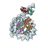
| Title | Structure of a single human ELF2 transcription factor in complex with a nucleosome | |||||||||||||||
 Map data Map data | RELION Refine3D output | |||||||||||||||
 Sample Sample |
| |||||||||||||||
 Keywords Keywords | nucleosome / transcription factor / DNA BINDING PROTEIN | |||||||||||||||
| Biological species |  Xenopus laevis (African clawed frog) / Xenopus laevis (African clawed frog) /  Homo sapiens (human) / synthetic construct (others) Homo sapiens (human) / synthetic construct (others) | |||||||||||||||
| Method | single particle reconstruction / cryo EM / Resolution: 3.1 Å | |||||||||||||||
 Authors Authors | Xiao T / Crowe-McAuliffe C / Dienemann C / Taipale J | |||||||||||||||
| Funding support |  United Kingdom, United Kingdom,  Germany, 4 items Germany, 4 items
| |||||||||||||||
 Citation Citation | Journal: To Be Published Title: The pioneer transcription factor ELF2 remodels the nucleosome near transcription start sites Authors: Xiao T / Crowe-McAuliffe C / Ochmann M / Du Y / Hou T / Chen Y / Zhu F / Seyednasrollah F / Osmala M / Vanharanta S / Morgunova E / Cramer P / Rogerson C / Dienemann C / Taipale J | |||||||||||||||
| History |
|
- Structure visualization
| Supplemental images |
|---|

- Downloads & links
-EMDB archive
| Map data | emd_58724.map.gz | 40.7 MB |  EMDB map data format EMDB map data format | |
|---|---|---|---|---|
| Header (meta data) | emd-58724-v30.xmlemd-58724.xml | 23.1 KB 23.1 KB | Display Display | EMDB header |
| FSC (resolution estimation) | emd_58724_fsc.xml | 11 KB | Display | FSC data file |
| Images |  emd_58724.png emd_58724.png | 91 KB | ||
| Masks | emd_58724_msk_1.map | 52.7 MB | Mask map | |
| Filedesc metadata | emd-58724.cif.gz | 6.2 KB | ||
| Others | emd_58724_half_map_1.map.gzemd_58724_half_map_2.map.gz | 40.8 MB 40.8 MB | ||
| Archive directory |  http://ftp.pdbj.org/pub/emdb/structures/EMD-58724ftp://ftp.pdbj.org/pub/emdb/structures/EMD-58724 http://ftp.pdbj.org/pub/emdb/structures/EMD-58724ftp://ftp.pdbj.org/pub/emdb/structures/EMD-58724 | HTTPS FTP |
-Related structure data


-Links
| EMDB pages | EMDB (EBI/PDBe) / EMDataResource |
|---|
-Map
| File | Download / File: emd_58724.map.gz / Format: CCP4 / Size: 52.7 MB / Type: IMAGE STORED AS FLOATING POINT NUMBER (4 BYTES) | ||||||||||||||||||||||||||||||||||||
|---|---|---|---|---|---|---|---|---|---|---|---|---|---|---|---|---|---|---|---|---|---|---|---|---|---|---|---|---|---|---|---|---|---|---|---|---|---|
| Annotation | RELION Refine3D output | ||||||||||||||||||||||||||||||||||||
| Projections & slices | Image control
Images are generated by Spider. | ||||||||||||||||||||||||||||||||||||
| Voxel size | X=Y=Z: 0.834 Å | ||||||||||||||||||||||||||||||||||||
| Density |
| ||||||||||||||||||||||||||||||||||||
| Symmetry | Space group: 1 | ||||||||||||||||||||||||||||||||||||
| Details | EMDB XML:
|
 Z (Sec.)
 Y (Row.)
 X (Col.)


-Supplemental data
-Mask #1
| File | emd_58724_msk_1.map | ||||||||||||
|---|---|---|---|---|---|---|---|---|---|---|---|---|---|
| Projections & Slices |
| ||||||||||||
| Density Histograms |


-Half map: Half map 1
| File | emd_58724_half_map_1.map | ||||||||||||
|---|---|---|---|---|---|---|---|---|---|---|---|---|---|
| Annotation | Half map 1 | ||||||||||||
| Projections & Slices |
| ||||||||||||
| Density Histograms |


-Half map: Half map 2
| File | emd_58724_half_map_2.map | ||||||||||||
|---|---|---|---|---|---|---|---|---|---|---|---|---|---|
| Annotation | Half map 2 | ||||||||||||
| Projections & Slices |
| ||||||||||||
| Density Histograms |


- Sample components
-Entire : Nucleosome in complex with a single ELF2 DNA-binding domain
| Entire | Name: Nucleosome in complex with a single ELF2 DNA-binding domain |
|---|---|
| Components |
|
-Supramolecule #1: Nucleosome in complex with a single ELF2 DNA-binding domain
| Supramolecule | Name: Nucleosome in complex with a single ELF2 DNA-binding domain type: complex / ID: 1 / Parent: 0 / Macromolecule list: all |
|---|---|
| Source (natural) | Organism: Xenopus laevis (African clawed frog) |
-Macromolecule #1: histone H3
| Macromolecule | Name: histone H3 / type: protein_or_peptide / ID: 1 / Number of copies: 2 / Enantiomer: LEVO |
|---|---|
| Source (natural) | Organism: Xenopus laevis (African clawed frog) |
| Molecular weight | Theoretical: 15.451192 KDa |
| Recombinant expression | Organism:   Escherichia coli BL21(DE3) (bacteria) Escherichia coli BL21(DE3) (bacteria) |
| Sequence | String: ARTKQTARKS TGGKAPRKQL ATKAARKSAP ATGGVKKPHR YRPGTVALRE IRRYQKSTEL LIRKLPFQRL VREIAQDFKT DLRFQSSAVM ALQEASEAYL VALFEDTNLC AIHAKRVTIM PKDIQLARRI RGERA |
-Macromolecule #2: Histone H4
| Macromolecule | Name: Histone H4 / type: protein_or_peptide / ID: 2 / Number of copies: 2 / Enantiomer: LEVO |
|---|---|
| Source (natural) | Organism: Xenopus laevis (African clawed frog) |
| Molecular weight | Theoretical: 11.394426 KDa |
| Recombinant expression | Organism: Escherichia coli BL21(DE3) (bacteria) |
| Sequence | String: SGRGKGGKGL GKGGAKRHRK VLRDNIQGIT KPAIRRLARR GGVKRISGLI YEETRGVLKV FLENVIRDAV TYTEHAKRK TVTAMDVVYA LKRQGRTLYG FGG |
-Macromolecule #3: Histone H2A type 1
| Macromolecule | Name: Histone H2A type 1 / type: protein_or_peptide / ID: 3 / Number of copies: 2 / Enantiomer: LEVO |
|---|---|
| Source (natural) | Organism: Xenopus laevis (African clawed frog) |
| Molecular weight | Theoretical: 14.109436 KDa |
| Recombinant expression | Organism: Escherichia coli BL21(DE3) (bacteria) |
| Sequence | String: SGRGKQGGKT RAKAKTRSSR AGLQFPVGRV HRLLRKGNYA ERVGAGAPVY LAAVLEYLTA EILELAGNAA RDNKKTRII PRHLQLAVRN DEELNKLLGR VTIAQGGVLP NIQSVLLPKK TESSKSAKSK |
-Macromolecule #4: Histone H2B 1.1
| Macromolecule | Name: Histone H2B 1.1 / type: protein_or_peptide / ID: 4 / Number of copies: 2 / Enantiomer: LEVO |
|---|---|
| Source (natural) | Organism: Xenopus laevis (African clawed frog) |
| Molecular weight | Theoretical: 13.655948 KDa |
| Recombinant expression | Organism: Escherichia coli BL21(DE3) (bacteria) |
| Sequence | String: AKSAPAPKKG SKKAVTKTQK KDGKKRRKTR KESYAIYVYK VLKQVHPDTG ISSKAMSIMN SFVNDVFERI AGEASRLAH YNKRSTITSR EIQTAVRLLL PGELAKHAVS EGTKAVTKYT SAK |
-Macromolecule #5: ETS-related transcription factor Elf-2
| Macromolecule | Name: ETS-related transcription factor Elf-2 / type: protein_or_peptide / ID: 5 / Number of copies: 2 / Enantiomer: LEVO |
|---|---|
| Source (natural) | Organism: Homo sapiens (human) |
| Molecular weight | Theoretical: 16.820217 KDa |
| Recombinant expression | Organism: Escherichia coli (E. coli) |
| Sequence | String: TQQSPISNGS PELGIKKKPR EGKGNTTYLW EFLLDLLQDK NTCPRYIKWT QREKGIFKLV DSKAVSKLWG KHKNKPDMNY ETMGRALRY YYQRGILAKV EGQRLVYQFK DMPKNIVVID DDKSETCNED LAGTTDEKSL ERVSLS |
-Macromolecule #6: DNA (147-MER)
| Macromolecule | Name: DNA (147-MER) / type: dna / ID: 6 / Number of copies: 1 / Classification: DNA |
|---|---|
| Source (natural) | Organism: synthetic construct (others) |
| Molecular weight | Theoretical: 45.288875 KDa |
| Sequence | String: CTGGAGAATC CCGGTCTGCA GGCCGCTCAA TTGGTCGTAG ACAGCTCTAG CACCGCTTAA ACGCACGTAC GCGCTGTCCC CCGCGTTTTA ACCGCCAAGG GGATTACTCC GGAAGCGTTA GTTGTATGTC AGATATATAC ATCCTGT |
-Macromolecule #7: DNA (147-MER)
| Macromolecule | Name: DNA (147-MER) / type: dna / ID: 7 / Number of copies: 1 / Classification: DNA |
|---|---|
| Source (natural) | Organism: synthetic construct (others) |
| Molecular weight | Theoretical: 45.45798 KDa |
| Sequence | String: ACAGGATGTA TATATCTGAC ATACAACTAA CGCTTCCGGA GTAATCCCCT TGGCGGTTAA AACGCGGGGG ACAGCGCGTA CGTGCGTTTA AGCGGTGCTA GAGCTGTCTA CGACCAATTG AGCGGCCTGC AGACCGGGAT TCTCCAG |
-Experimental details
-Structure determination
| Method | cryo EM |
|---|---|
 Processing Processing | single particle reconstruction |
| Aggregation state | particle |
-Sample preparation
| Buffer | pH: 7.5 |
|---|---|
| Grid | Model: Quantifoil R2/1 / Material: COPPER / Mesh: 200 / Pretreatment - Type: GLOW DISCHARGE |
| Vitrification | Cryogen name: ETHANE |
- Electron microscopy
| Microscope | TFS KRIOS |
|---|---|
| Specialist optics | Energy filter - Name: GIF Quantum LS / Energy filter - Slit width: 20 eV |
| Image recording | Film or detector model: GATAN K3 (6k x 4k) / Average electron dose: 40.0 e/Å2 |
| Electron beam | Acceleration voltage: 300 kV / Electron source:  FIELD EMISSION GUN FIELD EMISSION GUN |
| Electron optics | Illumination mode: FLOOD BEAM / Imaging mode: BRIGHT FIELD / Cs: 2.7 mm / Nominal defocus max: 2.0 µm / Nominal defocus min: 0.5 µm / Nominal magnification: 105000 |
| Sample stage | Cooling holder cryogen: NITROGEN |
| Experimental equipment |  Model: Titan Krios / Image courtesy: FEI Company |

