[English] 日本語
Yorodumi
- EMDB-58601: Cryo-EM structure of the CO dehydrogenase (CODH) subcomplex from ... -

+
Open data


ID or keywords:

Loading...

-
Basic information

Entry
Database: EMDB / ID: EMD-58601
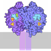
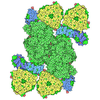
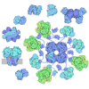
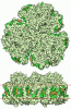
TitleCryo-EM structure of the CO dehydrogenase (CODH) subcomplex from Methanosarcina acetivorans
Map dataSharpened EM map of the CO dehydrogenase (CODH) subcomplex from a preparation of the acetyl-CoA decarbonylase/synthase (ACDS) complex from Methanosarcina acetivorans
Sample
  • Complex: CO dehydrogenase (CODH) isoform Cdh2
    • Protein or peptide: Acetyl-CoA decarbonylase/synthase complex subunit epsilon 2
    • Protein or peptide: Acetyl-CoA decarbonylase/synthase complex subunit alpha 2
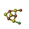
  • Ligand: IRON/SULFUR CLUSTER
  • Ligand: Fe(3)-Ni(1)-S(4) cluster
Keywordsmethanogenesis / acetyl-CoA / anaerobic metabolism / CO dehydrogenase / METAL BINDING PROTEIN
Function / homology
Function and homology information


methanogenesis, from acetate / nitric oxide catabolic process / anaerobic carbon monoxide dehydrogenase / hydroxylamine reductase activity / anaerobic carbon-monoxide dehydrogenase activity / acetyl-CoA metabolic process / nickel cation binding / response to hydrogen peroxide / peroxidase activity / 4 iron, 4 sulfur cluster binding / iron ion binding
Similarity search - Function
Acetyl-CoA decarbonylase/synthase complex subunit alpha / CO dehydrogenase beta subunit/acetyl-CoA synthase epsilon subunit / CO dehydrogenase beta subunit/acetyl-CoA synthase epsilon subunit / Hydroxylamine reductase/Ni-containing CO dehydrogenase / Prismane/CO dehydrogenase family / Prismane-like, alpha/beta-sandwich / Prismane-like superfamily / DHS-like NAD/FAD-binding domain superfamily / 4Fe-4S ferredoxin, iron-sulphur binding, conserved site / 4Fe-4S ferredoxin-type iron-sulfur binding region signature. ...Acetyl-CoA decarbonylase/synthase complex subunit alpha / CO dehydrogenase beta subunit/acetyl-CoA synthase epsilon subunit / CO dehydrogenase beta subunit/acetyl-CoA synthase epsilon subunit / Hydroxylamine reductase/Ni-containing CO dehydrogenase / Prismane/CO dehydrogenase family / Prismane-like, alpha/beta-sandwich / Prismane-like superfamily / DHS-like NAD/FAD-binding domain superfamily / 4Fe-4S ferredoxin, iron-sulphur binding, conserved site / 4Fe-4S ferredoxin-type iron-sulfur binding region signature. / 4Fe-4S ferredoxin-type iron-sulfur binding domain profile. / 4Fe-4S ferredoxin-type, iron-sulphur binding domain
Similarity search - Domain/homology
Acetyl-CoA decarbonylase/synthase complex subunit epsilon 2 / Acetyl-CoA decarbonylase/synthase complex subunit alpha 2
Similarity search - Component
Biological speciesMethanosarcina acetivorans (archaea)
Methodsingle particle reconstruction / cryo EM / Resolution: 3.55 Å
AuthorsZimmer E / Reif-Trauttmansdorff T / Schuller JM
Funding supportEuropean Union, Germany, 2 items
OrganizationGrant numberCountry
European Research Council (ERC)101075992European Union
German Research Foundation (DFG)RTG 2937 Germany
CitationJournal: bioRxiv / Year: 2026
Title: Interface swapping orchestrates carbon transfer in the archaeal acetyl-CoA decarbonylase/synthase.
Authors: Erik Zimmer / Tristan Reif-Trauttmansdorff / Anthony Ciancone / Sofia Appelgren / Jörg Kahnt / Darja Deobald / Frank Abendroth / Olalla Vázquez / Georg K A Hochberg / Francis J O'Reilly / Jan M Schuller /
Abstract: The Wood-Ljungdahl pathway is one of biology's most ancient routes for carbon fixation and energy metabolism, used by organisms such as methanogenic archaea. One of its central metabolic complexes is ...The Wood-Ljungdahl pathway is one of biology's most ancient routes for carbon fixation and energy metabolism, used by organisms such as methanogenic archaea. One of its central metabolic complexes is the acetyl-CoA decarbonylase/synthase (ACDS) complex, catalyzing acetyl-CoA synthesis and cleavage through the coordinated action of carbon monoxide dehydrogenase (CODH), acetyl-CoA synthase (ACS), and corrinoid iron-sulfur protein (CoFeSP). Unlike bacterial CODH/ACS, archaeal ACDS lacks a stable bifunctional CODH-ACS architecture, raising the question of how reactive CO and methyl intermediates are efficiently transferred between catalytic modules. Using cryo-electron microscopy, crosslinking mass spectrometry, small-angle X-ray scattering, and biophysical analyses, we resolved the organization and dynamics of the ~2 MDa archaeal ACDS supercomplex from . We identified CoFeSP as a central architectural scaffold that self-assembles into hexa- to octameric oligomers via a conserved N-terminal region of the CdhD subunit. This scaffold likely tethers CODH and ACS through conserved disordered terminal regions, positioning the catalytic modules in the complex's periphery. We propose a mechanism in which ACS transiently alternates between CODH and CoFeSP, enabling efficient CO and methyl-group transfer without stable binary complexes. This dynamic organization represents a fundamental difference to the stable bifunctional CODH/ACS in bacteria, highlighting how transient interactions enable efficient acetyl-CoA metabolism in archaea.
History
DepositionJun 16, 2026-
Header (metadata) releaseJul 29, 2026-
Map releaseJul 29, 2026-
UpdateJul 29, 2026-
Current statusJul 29, 2026Processing site: PDBe / Status: Released

-
Structure visualization

Supplemental images

Downloads & links

-
Map

FileDownload / File: emd_58601.map.gz / Format: CCP4 / Size: 178 MB / Type: IMAGE STORED AS FLOATING POINT NUMBER (4 BYTES)
AnnotationSharpened EM map of the CO dehydrogenase (CODH) subcomplex from a preparation of the acetyl-CoA decarbonylase/synthase (ACDS) complex from Methanosarcina acetivorans
Projections & slices

Image control

Size
Brightness
Contrast
Others
AxesZ (Sec.)Y (Row.)X (Col.)
0.85 Å/pix.
x 360 pix.
= 306. Å
0.85 Å/pix.
x 360 pix.
= 306. Å
0.85 Å/pix.
x 360 pix.
= 306. Å

Surface

Projections

Slices (1/3)

Slices (1/2)

Slices (2/3)

Images are generated by Spider.

Voxel sizeX=Y=Z: 0.85 Å
Density
Contour LevelBy AUTHOR: 0.13
Minimum - Maximum-0.7851446 - 1.823124
Average (Standard dev.)0.0008196895 (±0.023217909)
SymmetrySpace group: 1
Details

EMDB XML:

Map geometry
Axis orderXYZ
Origin000
Dimensions360360360
Spacing360360360
CellA=B=C: 306.0 Å
α=β=γ: 90.0 °

-
Supplemental data

-
Mask #1

Fileemd_58601_msk_1.map
Projections & Slices
AxesZYX

Projections

Slices (1/2)
Density Histograms

-
Half map: Half map B

Fileemd_58601_half_map_1.map
AnnotationHalf map B
Projections & Slices
AxesZYX

Projections

Slices (1/2)
Density Histograms

-
Half map: Half map A

Fileemd_58601_half_map_2.map
AnnotationHalf map A
Projections & Slices
AxesZYX

Projections

Slices (1/2)
Density Histograms

-
Sample components

-
Entire : CO dehydrogenase (CODH) isoform Cdh2

EntireName: CO dehydrogenase (CODH) isoform Cdh2
Components
  • Complex: CO dehydrogenase (CODH) isoform Cdh2
    • Protein or peptide: Acetyl-CoA decarbonylase/synthase complex subunit epsilon 2
    • Protein or peptide: Acetyl-CoA decarbonylase/synthase complex subunit alpha 2
  • Ligand: IRON/SULFUR CLUSTER
  • Ligand: Fe(3)-Ni(1)-S(4) cluster

-
Supramolecule #1: CO dehydrogenase (CODH) isoform Cdh2

SupramoleculeName: CO dehydrogenase (CODH) isoform Cdh2 / type: complex / ID: 1 / Parent: 0 / Macromolecule list: #1-#2
Source (natural)Organism: Methanosarcina acetivorans (archaea) / Strain: MCD31
Molecular weightTheoretical: 98 kDa/nm

-
Macromolecule #1: Acetyl-CoA decarbonylase/synthase complex subunit epsilon 2

MacromoleculeName: Acetyl-CoA decarbonylase/synthase complex subunit epsilon 2
type: protein_or_peptide / ID: 1 / Number of copies: 2 / Enantiomer: LEVO
Source (natural)Organism: Methanosarcina acetivorans (archaea) / Strain: MCD31
Molecular weightTheoretical: 18.487211 KDa
SequenceString:
MVDTTKNTKL FTSYGVTTSK TTTPEIAAKL ISKAKRPLLV VGTKVLDPEL LDRAVKIAQK ANIPIAATGS SMPGFVGKDV DAKYINLHQ LGFYVTDPNW PGLDGNGTYD TLIVLGHIKY YINQVLSGTK NFSTVKAIAI ERNYIQNATM SFGNLSKADH Y AALDELID AL

UniProtKB: Acetyl-CoA decarbonylase/synthase complex subunit epsilon 2

-
Macromolecule #2: Acetyl-CoA decarbonylase/synthase complex subunit alpha 2

MacromoleculeName: Acetyl-CoA decarbonylase/synthase complex subunit alpha 2
type: protein_or_peptide / ID: 2 / Number of copies: 2 / Enantiomer: LEVO / EC number: anaerobic carbon monoxide dehydrogenase
Source (natural)Organism: Methanosarcina acetivorans (archaea) / Strain: MCD31
Molecular weightTheoretical: 88.055109 KDa
SequenceString: MSKLTTGSFS IEDLESVQIT INNIVGAAKE AAEEKAKELG PMGPTAMAGL ASYRSWNLLL LDRYEPVLTP MCDQCCYCTY GPCDLSGNK RGACGIDMAG QTGREFFLRV ITGTACHAAH GRHLLDHVIE VFGEDLPLNL GESNVLTPNV TICTGLSPKT L GECRAPME ...String:
MSKLTTGSFS IEDLESVQIT INNIVGAAKE AAEEKAKELG PMGPTAMAGL ASYRSWNLLL LDRYEPVLTP MCDQCCYCTY GPCDLSGNK RGACGIDMAG QTGREFFLRV ITGTACHAAH GRHLLDHVIE VFGEDLPLNL GESNVLTPNV TICTGLSPKT L GECRAPME YVEEQLTQLL ATIHAGQESA EIDYDSKALF SGSLDHVGME VSDIAQVSAY DFPKADPEAP LIEIGMGSID KS KPLIVAI GHNVAGVTYI MDYMEENNLT DKMEIAGLCC TAFDMTRYKE ADRRAPYAKI VGSLAKELKV IRSGMPDVIV VDE QCVRGD VLSESQKLKI PVIASNEKIM MGLPDRTDAD VDSIVEEIKS GAIPGCVMLD YDKLGELIPK IAEVMAPIRD AEGI TAIPT DEEFKVYIDK CVKCGECMLA CPEELDIPEA LEYAAKGSYE YLEALHDVCI GCRRCEQVCK KEIPILNVLE KAAQK SISE EKGWVRSGRG QASDAEIRKE GLNLVMGTTP GIIAIIGCPN YPAGTKDVYL IAEEFLKRNY LLAVSGCSAM DIGMFK DED GKTLYEKYPG TFAGGGLLNT GSCVSNAHIS GAAEKVAGIF AQRTLAGNLA EIADYTLNRV GACGLAWGAY SQKAASI GT GCNIYGIPAV LGPHSSKYRR ALIAKNYDES KWKVYDGRDG SEMTIPPAPE FLLTTAETWQ EAIPMMAKAC IRPSDNNM G RSIKLTHWME LSKKYLGVEP EDWWKFVRNE ADLPLAKREE LLKRLEAEHG WEIDWKRKKI ISGPKIKFDV SAQPTNLKR LCKEA

UniProtKB: Acetyl-CoA decarbonylase/synthase complex subunit alpha 2

-
Macromolecule #3: IRON/SULFUR CLUSTER

MacromoleculeName: IRON/SULFUR CLUSTER / type: ligand / ID: 3 / Number of copies: 7 / Formula: SF4
Molecular weightTheoretical: 351.64 Da
Chemical component information

ChemComp-FS1:
IRON/SULFUR CLUSTER

-
Macromolecule #4: Fe(3)-Ni(1)-S(4) cluster

MacromoleculeName: Fe(3)-Ni(1)-S(4) cluster / type: ligand / ID: 4 / Number of copies: 2 / Formula: RQM
Molecular weightTheoretical: 410.333 Da
Chemical component information

ChemComp-RQM:
Fe(3)-Ni(1)-S(4) cluster

-
Experimental details

-
Structure determination

Methodcryo EM
Processingsingle particle reconstruction
Aggregation stateparticle

-
Sample preparation

Concentration1 mg/mL
BufferpH: 7.2
Component:
ConcentrationFormulaName
50.0 mMKH2PO4potassium dihydrogen phosphate
150.0 mMNaClsodium chloride

Details: buffer was anaerobised
GridModel: Quantifoil R1.2/1.3 / Material: COPPER / Pretreatment - Type: GLOW DISCHARGE / Pretreatment - Time: 25 sec.
Details: QUANTIFOIL R 1.2/1.3 copper grids were glow discharged for 25 s with a current of 15 mA in a PELCO easiGlow device (Ted Pella).
VitrificationCryogen name: ETHANE-PROPANE / Chamber humidity: 100 % / Chamber temperature: 277 K / Instrument: FEI VITROBOT MARK IV / Details: blotted with blot force 4 for 4 s.
Detailscrosslinked with 1 mM BS3; addition of 0.04 % (w/v) n-Octyl-beta-D-glucopyranoside

-
Electron microscopy

MicroscopeJEOL CRYO ARM 200
Image recordingFilm or detector model: GATAN K2 SUMMIT (4k x 4k) / Number grids imaged: 1 / Number real images: 5987 / Average electron dose: 50.0 e/Å2
Electron beamAcceleration voltage: 200 kV / Electron source: FIELD EMISSION GUN
Electron opticsIllumination mode: OTHER / Imaging mode: BRIGHT FIELD / Cs: 2.7 mm / Nominal defocus max: 2.5 µm / Nominal defocus min: 0.5 µm

+
Image processing

Particle selectionNumber selected: 371631
CTF correctionSoftware - Name: cryoSPARC (ver. 4) / Type: PHASE FLIPPING AND AMPLITUDE CORRECTION
Startup modelType of model: INSILICO MODEL
Final reconstructionApplied symmetry - Point group: C2 (2 fold cyclic) / Resolution.type: BY AUTHOR / Resolution: 3.55 Å / Resolution method: FSC 0.143 CUT-OFF / Software - Name: cryoSPARC (ver. 5) / Number images used: 187510
Initial angle assignmentType: PROJECTION MATCHING
Final angle assignmentType: MAXIMUM LIKELIHOOD
FSC plot (resolution estimation)

-
Atomic model buiding 1

Initial modelChain - Source name: AlphaFold / Chain - Initial model type: in silico model
SoftwareName: Coot (ver. 0.9.8.96)
RefinementSpace: REAL / Protocol: OTHER
Output model

PDB-31ox:
Cryo-EM structure of the CO dehydrogenase (CODH) subcomplex from Methanosarcina acetivorans

+
About Yorodumi

-
News

-
Feb 9, 2022. New format data for meta-information of EMDB entries

New format data for meta-information of EMDB entries

  • Version 3 of the EMDB header file is now the official format.
  • The previous official version 1.9 will be removed from the archive.

Related info.:EMDB header

External links:wwPDB to switch to version 3 of the EMDB data model

-
Aug 12, 2020. Covid-19 info

Covid-19 info

URL: https://pdbj.org/emnavi/covid19.php

New page: Covid-19 featured information page in EM Navigator.

Related info.:Covid-19 info / Mar 5, 2020. Novel coronavirus structure data

+
Mar 5, 2020. Novel coronavirus structure data

Novel coronavirus structure data

Related info.:Yorodumi Speices / Aug 12, 2020. Covid-19 info

External links:COVID-19 featured content - PDBj / Molecule of the Month (242):Coronavirus Proteases

+
Jan 31, 2019. EMDB accession codes are about to change! (news from PDBe EMDB page)

EMDB accession codes are about to change! (news from PDBe EMDB page)

  • The allocation of 4 digits for EMDB accession codes will soon come to an end. Whilst these codes will remain in use, new EMDB accession codes will include an additional digit and will expand incrementally as the available range of codes is exhausted. The current 4-digit format prefixed with “EMD-” (i.e. EMD-XXXX) will advance to a 5-digit format (i.e. EMD-XXXXX), and so on. It is currently estimated that the 4-digit codes will be depleted around Spring 2019, at which point the 5-digit format will come into force.
  • The EM Navigator/Yorodumi systems omit the EMD- prefix.

Related info.:Q: What is EMD? / ID/Accession-code notation in Yorodumi/EM Navigator

External links:EMDB Accession Codes are Changing Soon! / Contact to PDBj

+
Jul 12, 2017. Major update of PDB

Major update of PDB

  • wwPDB released updated PDB data conforming to the new PDBx/mmCIF dictionary.
  • This is a major update changing the version number from 4 to 5, and with Remediation, in which all the entries are updated.
  • In this update, many items about electron microscopy experimental information are reorganized (e.g. em_software).
  • Now, EM Navigator and Yorodumi are based on the updated data.

External links:wwPDB Remediation / Enriched Model Files Conforming to OneDep Data Standards Now Available in the PDB FTP Archive

-
Yorodumi

Thousand views of thousand structures

  • Yorodumi is a browser for structure data from EMDB, PDB, SASBDB, etc.
  • This page is also the successor to EM Navigator detail page, and also detail information page/front-end page for Omokage search.
  • The word "yorodu" (or yorozu) is an old Japanese word meaning "ten thousand". "mi" (miru) is to see.

Related info.:EMDB / PDB / SASBDB / Comparison of 3 databanks / Yorodumi Search / Aug 31, 2016. New EM Navigator & Yorodumi / Yorodumi Papers / Jmol/JSmol / Function and homology information / Changes in new EM Navigator and Yorodumi

Read more