Movie
Movie Controller

[English] 日本語
 Yorodumi
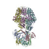
- EMDB-58342: P1a-state of wild type human mitochondrial LONP1 protease with bo... -
+ Open data

- Basic information
| Entry |  | |||||||||
|---|---|---|---|---|---|---|---|---|---|---|
| Title | P1a-state of wild type human mitochondrial LONP1 protease with bound substrate protein, ADP and aluminum fluoride | |||||||||
 Map data Map data | ||||||||||
 Sample Sample |
| |||||||||
 Keywords Keywords | AAA+ protease / Lon protease / transition state / MOTOR PROTEIN | |||||||||
| Function / homology |  Function and homology information Function and homology informationoxidation-dependent protein catabolic process / response to aluminum ion / PH domain binding / endopeptidase La / mitochondrial protein catabolic process / G-quadruplex DNA binding / ATP-dependent peptidase activity / protein quality control for misfolded or incompletely synthesized proteins / mitochondrial nucleoid / insulin receptor substrate binding ...oxidation-dependent protein catabolic process / response to aluminum ion / PH domain binding / endopeptidase La / mitochondrial protein catabolic process / G-quadruplex DNA binding / ATP-dependent peptidase activity / protein quality control for misfolded or incompletely synthesized proteins / mitochondrial nucleoid / insulin receptor substrate binding / Mitochondrial unfolded protein response (UPRmt) / chaperone-mediated protein complex assembly / DNA polymerase binding / response to hormone / negative regulation of insulin receptor signaling pathway / Mitochondrial protein degradation / : / mitochondrion organization / ADP binding / single-stranded DNA binding / cellular response to oxidative stress / sequence-specific DNA binding / response to hypoxia / single-stranded RNA binding / mitochondrial matrix / serine-type endopeptidase activity / ATP hydrolysis activity / mitochondrion / nucleoplasm / ATP binding / membrane / identical protein binding / cytosol Similarity search - Function | |||||||||
| Biological species |  Homo sapiens (human) / Homo sapiens (human) /  | |||||||||
| Method | single particle reconstruction / cryo EM / Resolution: 2.54 Å | |||||||||
 Authors Authors | Schenck N / Filipcik P / Abrahams JP | |||||||||
| Funding support |  Switzerland, 1 items Switzerland, 1 items
| |||||||||
 Citation Citation | Journal: biorxiv Title: Human mitochondrial Lon protease initiates unidirectional degradation from either substrate terminus Authors: Schenck N / Filipcik P / Abrahams JP | |||||||||
| History |
|
- Structure visualization
| Supplemental images |
|---|

- Downloads & links
-EMDB archive
| Map data | emd_58342.map.gz | 256.2 MB | EMDB map data format | |
|---|---|---|---|---|
| Header (meta data) | emd-58342-v30.xmlemd-58342.xml | 18.5 KB 18.5 KB | Display Display | EMDB header |
| FSC (resolution estimation) | emd_58342_fsc.xml | 19.1 KB | Display | FSC data file |
| Images |  emd_58342.png emd_58342.png | 104.8 KB | ||
| Filedesc metadata | emd-58342.cif.gz | 6.5 KB | ||
| Others | emd_58342_half_map_1.map.gzemd_58342_half_map_2.map.gz | 475.6 MB 475.6 MB | ||
| Archive directory |  http://ftp.pdbj.org/pub/emdb/structures/EMD-58342ftp://ftp.pdbj.org/pub/emdb/structures/EMD-58342 http://ftp.pdbj.org/pub/emdb/structures/EMD-58342ftp://ftp.pdbj.org/pub/emdb/structures/EMD-58342 | HTTPS FTP |
-Related structure data
| Related structure data |  31epMC M: atomic model generated by this map C: citing same article ( |
|---|---|
| Similar structure data |
-Links
| EMDB pages | EMDB (EBI/PDBe) / EMDataResource |
|---|---|
| Related items in Molecule of the Month |

-Map
| File | Download / File: emd_58342.map.gz / Format: CCP4 / Size: 512 MB / Type: IMAGE STORED AS FLOATING POINT NUMBER (4 BYTES) | ||||||||||||||||||||||||||||||||||||
|---|---|---|---|---|---|---|---|---|---|---|---|---|---|---|---|---|---|---|---|---|---|---|---|---|---|---|---|---|---|---|---|---|---|---|---|---|---|
| Projections & slices | Image control
Images are generated by Spider. | ||||||||||||||||||||||||||||||||||||
| Voxel size | X=Y=Z: 0.73 Å | ||||||||||||||||||||||||||||||||||||
| Density |
| ||||||||||||||||||||||||||||||||||||
| Symmetry | Space group: 1 | ||||||||||||||||||||||||||||||||||||
| Details | EMDB XML:
|
 Z (Sec.)
 Y (Row.)
 X (Col.)


-Supplemental data
-Half map: #2
| File | emd_58342_half_map_1.map | ||||||||||||
|---|---|---|---|---|---|---|---|---|---|---|---|---|---|
| Projections & Slices |
| ||||||||||||
| Density Histograms |


-Half map: #1
| File | emd_58342_half_map_2.map | ||||||||||||
|---|---|---|---|---|---|---|---|---|---|---|---|---|---|
| Projections & Slices |
| ||||||||||||
| Density Histograms |


- Sample components
-Entire : Lon protease homolog, mitochondrial
| Entire | Name: Lon protease homolog, mitochondrial |
|---|---|
| Components |
|
-Supramolecule #1: Lon protease homolog, mitochondrial
| Supramolecule | Name: Lon protease homolog, mitochondrial / type: complex / ID: 1 / Parent: 0 / Macromolecule list: #2 Details: P1a-state of wild type human mitochondrial LONP1 protease with bound substrate protein, ADP and aluminum fluoride |
|---|---|
| Source (natural) | Organism: Homo sapiens (human) |
| Molecular weight | Theoretical: 585 KDa |
-Macromolecule #1: Lon protease homolog, mitochondrial
| Macromolecule | Name: Lon protease homolog, mitochondrial / type: protein_or_peptide / ID: 1 / Number of copies: 6 / Enantiomer: LEVO / EC number: endopeptidase La |
|---|---|
| Source (natural) | Organism: Homo sapiens (human) |
| Molecular weight | Theoretical: 96.419953 KDa |
| Recombinant expression | Organism: |
| Sequence | String: MHHHHHHGSM TIPDVFPHLP LIAITRNPVF PRFIKIIEVK NKKLVELLRR KVRLAQPYVG VFLKRDDSNE SDVVESLDEI YHTGTFAQI HEMQDLGDKL RMIVMGHRRV HISRQLEVEP EEPEAENKHK PRRKSKRGKK EAEDELSARH PAELAMEPTP E LPAEVLMV ...String: MHHHHHHGSM TIPDVFPHLP LIAITRNPVF PRFIKIIEVK NKKLVELLRR KVRLAQPYVG VFLKRDDSNE SDVVESLDEI YHTGTFAQI HEMQDLGDKL RMIVMGHRRV HISRQLEVEP EEPEAENKHK PRRKSKRGKK EAEDELSARH PAELAMEPTP E LPAEVLMV EVENVVHEDF QVTEEVKALT AEIVKTIRDI IALNPLYRES VLQMMQAGQR VVDNPIYLSD MGAALTGAES HE LQDVLEE TNIPKRLYKA LSLLKKEFEL SKLQQRLGRE VEEKIKQTHR KYLLQEQLKI IKKELGLEKD DKDAIEEKFR ERL KELVVP KHVMDVVDEE LSKLGLLDNH SSEFNVTRNY LDWLTSIPWG KYSNENLDLA RAQAVLEEDH YGMEDVKKRI LEFI AVSQL RGSTQGKILC FYGPPGVGKT SIARSIARAL NREYFRFSVG GMTDVAEIKG HRRTYVGAMP GKIIQCLKKT KTENP LILI DEVDKIGRGY QGDPSSALLE LLDPEQNANF LDHYLDVPVD LSKVLFICTA NVTDTIPEPL RDRMEMINVS GYVAQE KLA IAERYLVPQA RALCGLDESK AKLSSDVLTL LIKQYCRESG VRNLQKQVEK VLRKSAYKIV SGEAESVEVT PENLQDF VG KPVFTVERMY DVTPPGVVMG LAWTAMGGST LFVETSLRRP QDKDAKGDKD GSLEVTGQLG EVMKESARIA YTFARAFL M QHAPANDYLV TSHIHLHVPE GATPKDGPSA GCTIVTALLS LAMGRPVRQN LAMTGEVSLT GKILPVGGIK EKTIAAKRA GVTCIVLPAE NKKDFYDLAA FITEGLEVHF VEHYREIFDI AFPDEQAEAL AVER UniProtKB: Lon protease homolog, mitochondrial |
-Macromolecule #2: unknown translocated polypeptide substrate
| Macromolecule | Name: unknown translocated polypeptide substrate / type: protein_or_peptide / ID: 2 / Number of copies: 2 / Enantiomer: LEVO |
|---|---|
| Source (natural) | Organism: |
| Molecular weight | Theoretical: 1.124378 KDa |
| Recombinant expression | Organism: |
| Sequence | String: (UNK)(UNK)(UNK)(UNK)(UNK)(UNK)(UNK)(UNK)(UNK)(UNK) (UNK)(UNK)(UNK) |
-Macromolecule #3: ADENOSINE-5'-DIPHOSPHATE
| Macromolecule | Name: ADENOSINE-5'-DIPHOSPHATE / type: ligand / ID: 3 / Number of copies: 6 / Formula: ADP |
|---|---|
| Molecular weight | Theoretical: 427.201 Da |
| Chemical component information |  ChemComp-ADP: |
-Macromolecule #4: MAGNESIUM ION
| Macromolecule | Name: MAGNESIUM ION / type: ligand / ID: 4 / Number of copies: 4 / Formula: MG |
|---|---|
| Molecular weight | Theoretical: 24.305 Da |
-Macromolecule #5: ALUMINUM FLUORIDE
| Macromolecule | Name: ALUMINUM FLUORIDE / type: ligand / ID: 5 / Number of copies: 4 / Formula: AF3 |
|---|---|
| Molecular weight | Theoretical: 83.977 Da |
| Chemical component information |  ChemComp-AF3: |
-Experimental details
-Structure determination
| Method | cryo EM |
|---|---|
 Processing Processing | single particle reconstruction |
| Aggregation state | particle |
-Sample preparation
| Buffer | pH: 7.5 Details: Pure LonP1 fractions (1.0 mg/mL) were incubated with 1 mM ADP in SEC buffer (50 mM HEPES, 150 mM NaCl, 5 mM MgCl2, pH 7.5) for 20 min at room temperature. Subsequently, 3.125 mM NaF was ...Details: Pure LonP1 fractions (1.0 mg/mL) were incubated with 1 mM ADP in SEC buffer (50 mM HEPES, 150 mM NaCl, 5 mM MgCl2, pH 7.5) for 20 min at room temperature. Subsequently, 3.125 mM NaF was added, followed by 0.625 mM AlCl3 after additional 20 min incubation for each step to generate the LonP1-ADP-AlF3 complex. |
|---|---|
| Grid | Model: Quantifoil R1.2/1.3 / Material: COPPER / Pretreatment - Type: GLOW DISCHARGE / Pretreatment - Time: 60 sec. |
| Vitrification | Cryogen name: ETHANE |
- Electron microscopy
| Microscope | TFS KRIOS |
|---|---|
| Image recording | Film or detector model: FEI FALCON IV (4k x 4k) / Average electron dose: 40.5 e/Å2 |
| Electron beam | Acceleration voltage: 300 kV / Electron source:  FIELD EMISSION GUN FIELD EMISSION GUN |
| Electron optics | Illumination mode: FLOOD BEAM / Imaging mode: BRIGHT FIELD / Nominal defocus max: 2.0 µm / Nominal defocus min: 0.8 µm |
| Experimental equipment |  Model: Titan Krios / Image courtesy: FEI Company |