 Movie
Movie Controller

[English] 日本語
 Yorodumi
- EMDB-57826: In situ cryo-electron tomogram of phagophore expansion in Atg2-PM... -
+ Open data

- Basic information
| Entry |  | |||||||||||||||
|---|---|---|---|---|---|---|---|---|---|---|---|---|---|---|---|---|
| Title | In situ cryo-electron tomogram of phagophore expansion in Atg2-PM4 mutant S. cerevisiae #3 | |||||||||||||||
 Map data Map data | phagophore in direct contact with the phospholipid monolayer at the surface of a lipid droplet | |||||||||||||||
 Sample Sample |
| |||||||||||||||
 Keywords Keywords | Atg2 / phagophore expansion / autophagosome biogenesis / LIPID TRANSPORT | |||||||||||||||
| Biological species |   Saccharomyces cerevisiae BY4741 (yeast) Saccharomyces cerevisiae BY4741 (yeast) | |||||||||||||||
| Method | electron tomography / cryo EM | |||||||||||||||
 Authors Authors | Ortmann de Percin Northumberland C / Licheva M / Dabrowski R / Gomez-Sanchez R / Berkamp S / Schonnenbeck P / Graef M / Kraft C / Sachse C | |||||||||||||||
| Funding support |  Germany, European Union, 4 items Germany, European Union, 4 items
| |||||||||||||||
 Citation Citation | Journal: J Cell Sci / Year: 2026 Title: In situ visualization of autophagy reveals the presence of vesicles at the expanding phagophore rim. Authors: Claire Ortmann de Percin Northumberland / Mariya Licheva / Rahel Dabrowski / Rubén Gómez-Sánchez / Sabrina Berkamp / Philipp Schönnenbeck / Martin Graef / Claudine Kraft / Carsten Sachse /   Abstract: The autophagy core machinery mediates the enclosure of cytosolic cargo destined for degradation in the lysosome. The Atg9-Atg2-Atg18 complex coordinates phagophore expansion via directed lipid ...The autophagy core machinery mediates the enclosure of cytosolic cargo destined for degradation in the lysosome. The Atg9-Atg2-Atg18 complex coordinates phagophore expansion via directed lipid transfer until closure of the phagophore rim. Using an Atg2 variant (Atg2-PM4) as a model of decelerated autophagosome biogenesis, we visualized the morphological states prior to autophagosome closure by cryogenic correlative light and electron microscopy in S. cerevisiae. Using in situ cryo-electron tomography, we find an enlarged rim morphology of an expanding phagophore in Atg2-PM4 cells in comparison with Atg2 wildtype condition. Analysis of segmented rim membrane features reveal surrounding and membrane-attached vesicles. The dimensions of the enlarged rims are consistent with fusion of cytosolic vesicles with the growing phagophore. High-resolution imaging in this study suggests that, apart from the initial nucleation phase, vesicle fusion can also contribute to phagophore expansion during later stages of autophagosome biogenesis. | |||||||||||||||
| History |
|
- Structure visualization
| Supplemental images |
|---|

- Downloads & links
-EMDB archive
| Map data | emd_57826.map.gz | 2.3 GB |  EMDB map data format EMDB map data format | |
|---|---|---|---|---|
| Header (meta data) | emd-57826-v30.xmlemd-57826.xml | 15.4 KB 15.4 KB | Display Display | EMDB header |
| Images |  emd_57826.png emd_57826.png | 120.2 KB | ||
| Filedesc metadata | emd-57826.cif.gz | 4.7 KB | ||
| Archive directory |  https://data.pdbj.org/pub/emdb/structures/EMD-57826ftp://data.pdbj.org/pub/emdb/structures/EMD-57826 https://data.pdbj.org/pub/emdb/structures/EMD-57826ftp://data.pdbj.org/pub/emdb/structures/EMD-57826 | HTTPS FTP |
-Related structure data


-Links
| EMDB pages | EMDB (EBI/PDBe) / EMDataResource |
|---|
-Map
| File | Download / File: emd_57826.map.gz / Format: CCP4 / Size: 2.5 GB / Type: IMAGE STORED AS FLOATING POINT NUMBER (4 BYTES) | ||||||||||||||||||||||||||||||||
|---|---|---|---|---|---|---|---|---|---|---|---|---|---|---|---|---|---|---|---|---|---|---|---|---|---|---|---|---|---|---|---|---|---|
| Annotation | phagophore in direct contact with the phospholipid monolayer at the surface of a lipid droplet | ||||||||||||||||||||||||||||||||
| Projections & slices | Image control
Images are generated by Spider. generated in cubic-lattice coordinate | ||||||||||||||||||||||||||||||||
| Voxel size | X=Y=Z: 13.95 Å | ||||||||||||||||||||||||||||||||
| Density |
| ||||||||||||||||||||||||||||||||
| Symmetry | Space group: 1 | ||||||||||||||||||||||||||||||||
| Details | EMDB XML:
|
 Z (Sec.)
 Y (Row.)
 X (Col.)


-Supplemental data
- Sample components
-Entire : Atg2-PM4-3xmScarlet mutant Atg9-3xmTagBFP2 mutant both expressed ...
| Entire | Name: Atg2-PM4-3xmScarlet mutant Atg9-3xmTagBFP2 mutant both expressed under native promoters from plasmids while the native gene copies were knocked out |
|---|---|
| Components |
|
-Supramolecule #1: Atg2-PM4-3xmScarlet mutant Atg9-3xmTagBFP2 mutant both expressed ...
| Supramolecule | Name: Atg2-PM4-3xmScarlet mutant Atg9-3xmTagBFP2 mutant both expressed under native promoters from plasmids while the native gene copies were knocked out type: cell / ID: 1 / Parent: 0 Details: Atg2-PM4 mutant protein previously generated and characterised (doi:10.1083/jcb.201710116) |
|---|---|
| Source (natural) | Organism: Saccharomyces cerevisiae BY4741 (yeast) |
-Experimental details
-Structure determination
| Method | cryo EM |
|---|---|
 Processing Processing | electron tomography |
| Aggregation state | cell |
-Sample preparation
| Buffer | pH: 7 |
|---|---|
| Grid | Model: Quantifoil R2/1 / Material: COPPER / Mesh: 200 / Support film - Material: SILICON DIOXIDE / Support film - topology: HOLEY / Pretreatment - Type: PLASMA CLEANING / Pretreatment - Atmosphere: AIR |
| Vitrification | Cryogen name: ETHANE-PROPANE / Chamber humidity: 80 % / Chamber temperature: 303 K / Instrument: FEI VITROBOT MARK IV |
| Cryo protectant | none |
| Sectioning | Focused ion beam - Instrument: OTHER / Focused ion beam - Ion: OTHER / Focused ion beam - Voltage: 30 / Focused ion beam - Current: 0.01 / Focused ion beam - Duration: 10 / Focused ion beam - Temperature: 77 K / Focused ion beam - Initial thickness: 600 / Focused ion beam - Final thickness: 280 Focused ion beam - Details: polishing was done manually so duration is an approximation. The value given for _em_focused_ion_beam.instrument is TFS Aquilos 2. This is not in a list of allowed values ...Focused ion beam - Details: polishing was done manually so duration is an approximation. The value given for _em_focused_ion_beam.instrument is TFS Aquilos 2. This is not in a list of allowed values {'OTHER', 'DB235'} so OTHER is written into the XML file. |
| Fiducial marker | Manufacturer: Thermo Fisher Scientific / Diameter: 100 nm |
- Electron microscopy
| Microscope | FEI TALOS ARCTICA |
|---|---|
| Specialist optics | Energy filter - Name: GIF Bioquantum / Energy filter - Slit width: 30 eV |
| Image recording | Film or detector model: GATAN K3 BIOQUANTUM (6k x 4k) / Average electron dose: 2.16 e/Å2 |
| Electron beam | Acceleration voltage: 200 kV / Electron source:  FIELD EMISSION GUN FIELD EMISSION GUN |
| Electron optics | C2 aperture diameter: 100.0 µm / Illumination mode: FLOOD BEAM / Imaging mode: BRIGHT FIELD / Cs: 2.7 mm / Nominal defocus max: 5.0 µm / Nominal defocus min: 5.0 µm / Nominal magnification: 24000 |
| Sample stage | Specimen holder model: FEI TITAN KRIOS AUTOGRID HOLDER / Cooling holder cryogen: NITROGEN |
| Experimental equipment | 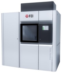 Model: Talos Arctica / Image courtesy: FEI Company |
-Image processing
| Final reconstruction | Algorithm: BACK PROJECTION / Software - Name: AreTomo (ver. 1) / Number images used: 60 |
|---|---|
| CTF correction | Software - Name: Warp / Type: PHASE FLIPPING AND AMPLITUDE CORRECTION |
