 Movie
Movie Controller


+ Open data

- Basic information
| Entry | Database: EMDB / ID: EMD-5720 | |||||||||
|---|---|---|---|---|---|---|---|---|---|---|
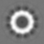
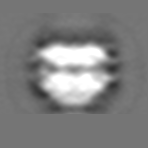
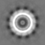
| Title | Structure of the Yersinia enterocolitica secretin YscC | |||||||||
 Map data Map data | Reconstruction of YscC secretin | |||||||||
 Sample Sample |
| |||||||||
 Keywords Keywords | type III secretion system / Yersinia enterocolitica / Gram-negative bacteria / YscC / secretin / cryo-electron microscopy / outer membrane | |||||||||
| Function / homology |  Function and homology information Function and homology informationtype III protein secretion system complex / type II protein secretion system complex / protein secretion by the type III secretion system / cell outer membrane / identical protein binding Similarity search - Function | |||||||||
| Biological species |  Yersinia enterocolitica (bacteria) Yersinia enterocolitica (bacteria) | |||||||||
| Method | single particle reconstruction / cryo EM / Resolution: 15.0 Å | |||||||||
 Authors Authors | Kowal J / Chami M / Ringler P / Muller AS / Kudryashev M / Castano-Diez D / Amstutz M / Cornelis GR / Stahlberg H / Engel A | |||||||||
 Citation Citation | Journal: Structure / Year: 2013 Title: Structure of the dodecameric Yersinia enterocolitica secretin YscC and its trypsin-resistant core. Authors: Julia Kowal / Mohamed Chami / Philippe Ringler / Shirley A Müller / Mikhail Kudryashev / Daniel Castaño-Díez / Marlise Amstutz / Guy R Cornelis / Henning Stahlberg / Andreas Engel /  Abstract: The type III secretion system machinery, also known as the injectisome, delivers bacterial effector proteins into eukaryotic cells during infection. The outer membrane YscC secretin is a major part ...The type III secretion system machinery, also known as the injectisome, delivers bacterial effector proteins into eukaryotic cells during infection. The outer membrane YscC secretin is a major part of Yersinia enterocolitica's injectisome and is among the first components to assemble, solely assisted by its pilotin, YscW. We have determined the three-dimensional structures of the native complex and its protease-resistant core to 12 Å resolution by cryo-electron microscopy (cryo-EM) and show that YscC forms a dodecameric complex. Cryo-EM of YscC reconstituted into proteoliposomes defines the secretin's membrane-spanning region. Native YscC consists of an outer membrane ring connected via a thin cylindrical wall to a conical, periplasmic region that exposes N-terminal petals connected by flexible linkers. These petals harbor the binding site of YscD, a component of the inner membrane ring. A change in their orientation adapts the length of the YscC secretin and facilitates its interaction with YscD. | |||||||||
| History |
|
- Structure visualization
| Movie |
Movie viewer |
|---|---|
| Structure viewer | EM map: SurfViewMolmilJmol/JSmol |
| Supplemental images |

- Downloads & links
-EMDB archive
| Map data | emd_5720.map.gz | 7.9 MB | EMDB map data format | |
|---|---|---|---|---|
| Header (meta data) | emd-5720-v30.xmlemd-5720.xml | 11.1 KB 11.1 KB | Display Display | EMDB header |
| Images | 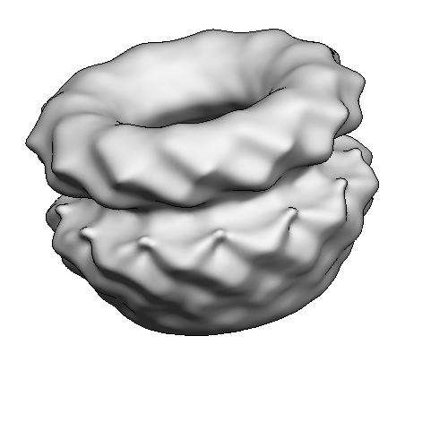 emd_5720.png emd_5720.png | 59.4 KB | ||
| Archive directory |  http://ftp.pdbj.org/pub/emdb/structures/EMD-5720ftp://ftp.pdbj.org/pub/emdb/structures/EMD-5720 http://ftp.pdbj.org/pub/emdb/structures/EMD-5720ftp://ftp.pdbj.org/pub/emdb/structures/EMD-5720 | HTTPS FTP |
-Related structure data


-Links
| EMDB pages | EMDB (EBI/PDBe) / EMDataResource |
|---|---|
| Related items in Molecule of the Month |

-Map
| File | Download / File: emd_5720.map.gz / Format: CCP4 / Size: 12.1 MB / Type: IMAGE STORED AS FLOATING POINT NUMBER (4 BYTES) | ||||||||||||||||||||||||||||||||||||||||||||||||||||||||||||||||||||
|---|---|---|---|---|---|---|---|---|---|---|---|---|---|---|---|---|---|---|---|---|---|---|---|---|---|---|---|---|---|---|---|---|---|---|---|---|---|---|---|---|---|---|---|---|---|---|---|---|---|---|---|---|---|---|---|---|---|---|---|---|---|---|---|---|---|---|---|---|---|
| Annotation | Reconstruction of YscC secretin | ||||||||||||||||||||||||||||||||||||||||||||||||||||||||||||||||||||
| Projections & slices | Image control
Images are generated by Spider. | ||||||||||||||||||||||||||||||||||||||||||||||||||||||||||||||||||||
| Voxel size | X=Y=Z: 1.95 Å | ||||||||||||||||||||||||||||||||||||||||||||||||||||||||||||||||||||
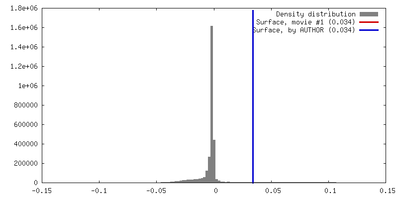
| Density |
| ||||||||||||||||||||||||||||||||||||||||||||||||||||||||||||||||||||
| Symmetry | Space group: 1 | ||||||||||||||||||||||||||||||||||||||||||||||||||||||||||||||||||||
| Details | EMDB XML:
CCP4 map header:
| ||||||||||||||||||||||||||||||||||||||||||||||||||||||||||||||||||||
 Z (Sec.)
 Y (Row.)
 X (Col.)


-Supplemental data
- Sample components
-Entire : YscC secretin from Yersinia enterocolitica
| Entire | Name: YscC secretin from Yersinia enterocolitica |
|---|---|
| Components |
|
-Supramolecule #1000: YscC secretin from Yersinia enterocolitica
| Supramolecule | Name: YscC secretin from Yersinia enterocolitica / type: sample / ID: 1000 / Details: Sample contained detergent DDM / Oligomeric state: 12 / Number unique components: 1 |
|---|---|
| Molecular weight | Experimental: 900 KDa / Theoretical: 768 KDa / Method: Gel filtration, Superose 6 30/100 GL column |
-Macromolecule #1: YscC secretin
| Macromolecule | Name: YscC secretin / type: protein_or_peptide / ID: 1 / Oligomeric state: Dodecamer / Recombinant expression: Yes |
|---|---|
| Source (natural) | Organism: Yersinia enterocolitica (bacteria) / Strain: W22703 / synonym: Yersinia / Location in cell: outer membrane |
| Molecular weight | Theoretical: 768 KDa |
| Recombinant expression | Organism: Yersinia enterocolitica (bacteria) / Recombinant strain: W22703 / Recombinant plasmid: pMA6, pRS6 |
| Sequence | UniProtKB: Type 3 secretion system secretin |
-Experimental details
-Structure determination
| Method | cryo EM |
|---|---|
 Processing Processing | single particle reconstruction |
| Aggregation state | particle |
-Sample preparation
| Concentration | 0.2 mg/mL |
|---|---|
| Buffer | pH: 7.8 Details: 10 mM Tris-HCl, 100 mM NaCl, 0.1 mM EDTA, 0.04% DDM |
| Grid | Details: 200 mesh Cu grid, covered with a very thin additional continuous carbon film, glow-discharged |
| Vitrification | Cryogen name: ETHANE / Chamber humidity: 95 % / Instrument: FEI VITROBOT MARK IV / Method: Plunging immediately after blotting. |
- Electron microscopy
| Microscope | FEI/PHILIPS CM200FEG |
|---|---|
| Date | Oct 1, 2010 |
| Image recording | Category: FILM / Film or detector model: KODAK SO-163 FILM / Digitization - Scanner: PRIMESCAN / Digitization - Sampling interval: 5 µm / Number real images: 100 / Average electron dose: 20 e/Å2 |
| Electron beam | Acceleration voltage: 200 kV / Electron source:  FIELD EMISSION GUN FIELD EMISSION GUN |
| Electron optics | Illumination mode: FLOOD BEAM / Imaging mode: BRIGHT FIELD / Cs: 2 mm / Nominal defocus max: 3.5 µm / Nominal defocus min: 1.0 µm / Nominal magnification: 50000 |
| Sample stage | Specimen holder model: GATAN LIQUID NITROGEN |
-Image processing
| Details | Particles were selected using Boxer/EMAN1.9. Image processing was done using EMAN2. |
|---|---|
| CTF correction | Details: each particle |
| Final reconstruction | Algorithm: OTHER / Resolution.type: BY AUTHOR / Resolution: 15.0 Å / Resolution method: FSC 0.5 CUT-OFF / Software - Name: EMAN2 Details: Mask parameters were optimized to minimize the influence of a flexible N-terminal domain that impaired projection alignment. Number images used: 35000 |
| Final two d classification | Number classes: 240 |
