 Movie
Movie Controller

[English] 日本語
 Yorodumi
- EMDB-5711: Novel Structural Labeling Method using Cryo-electron Tomography a... -
+ Open data

- Basic information
| Entry | Database: EMDB / ID: EMD-5711 | |||||||||
|---|---|---|---|---|---|---|---|---|---|---|
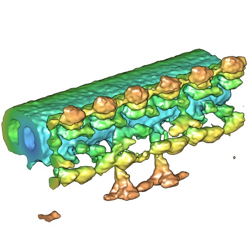
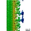
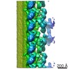
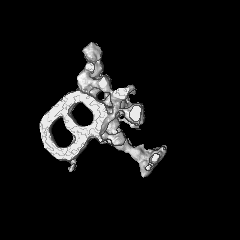
| Title | Novel Structural Labeling Method using Cryo-electron Tomography and Biotin-Streptavidin System | |||||||||
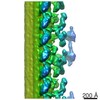
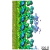
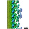
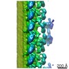
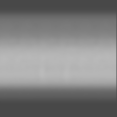
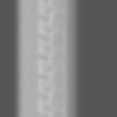
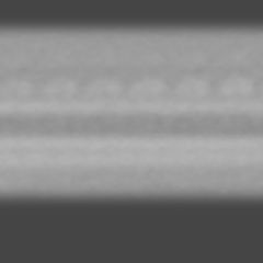
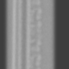
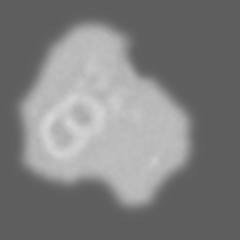
 Map data Map data | Averaged subtomogram of Chlamydomonas axoneme, labeled with Streptavidin | |||||||||
 Sample Sample |
| |||||||||
 Keywords Keywords | Cryo-electron tomography / Chlamydomonas reinhardtii / biotin-streptavidin / cilia and flagella / dynein / structural labeling | |||||||||
| Biological species |   Chlamydomonas reinhardtii (plant) Chlamydomonas reinhardtii (plant) | |||||||||
| Method | subtomogram averaging / cryo EM / Resolution: 57.0 Å | |||||||||
 Authors Authors | Oda T / Kikkawa M | |||||||||
 Citation Citation | Journal: J Struct Biol / Year: 2013 Title: Novel structural labeling method using cryo-electron tomography and biotin-streptavidin system. Authors: Toshiyuki Oda / Masahide Kikkawa /  Abstract: There are a number of large macromolecular complexes that play important roles in the cell, and identifying the positions of their components is a key step to understanding their structure and ...There are a number of large macromolecular complexes that play important roles in the cell, and identifying the positions of their components is a key step to understanding their structure and function. Several structural labeling methods have been applied to electron microscopy in order to locate a specific component within a macromolecular complex, but each method is associated with problems in specificity, occupancy, signal intensity or precision. Here, we report a novel method for identifying the 3D locations of proteins using biotin-streptavidin labeling and cryo-electron tomography. We labeled a biotinylation-tagged intermediate chain of an axonemal dynein by streptavidin within the Chlamydomonas axoneme and visualized the 3D positions of the labels using subtomogram averaging. Increase of the density attributed to the bound streptavidin was validated by Student's t-test. In conclusion, the combination of the biotin-streptavidin system and cryo-electron tomography is a powerful method to investigate the structure of large macromolecular complexes. | |||||||||
| History |
|
- Structure visualization
| Movie |
 Movie viewer Movie viewer |
|---|---|
| Structure viewer | EM map: SurfViewMolmilJmol/JSmol |
| Supplemental images |
- Downloads & links
-EMDB archive
| Map data | emd_5711.map.gz | 42.3 MB | EMDB map data format | |
|---|---|---|---|---|
| Header (meta data) | emd-5711-v30.xmlemd-5711.xml | 8.4 KB 8.4 KB | Display Display | EMDB header |
| Images | emd_5711_1.tif | 222.6 KB | ||
| Archive directory |  http://ftp.pdbj.org/pub/emdb/structures/EMD-5711ftp://ftp.pdbj.org/pub/emdb/structures/EMD-5711 http://ftp.pdbj.org/pub/emdb/structures/EMD-5711ftp://ftp.pdbj.org/pub/emdb/structures/EMD-5711 | HTTPS FTP |
-Related structure data
-Links
| EMDB pages | EMDB (EBI/PDBe) / EMDataResource |
|---|
-Map
| File | Download / File: emd_5711.map.gz / Format: CCP4 / Size: 51.5 MB / Type: IMAGE STORED AS FLOATING POINT NUMBER (4 BYTES) | ||||||||||||||||||||||||||||||||||||||||||||||||||||||||||||||||||||
|---|---|---|---|---|---|---|---|---|---|---|---|---|---|---|---|---|---|---|---|---|---|---|---|---|---|---|---|---|---|---|---|---|---|---|---|---|---|---|---|---|---|---|---|---|---|---|---|---|---|---|---|---|---|---|---|---|---|---|---|---|---|---|---|---|---|---|---|---|---|
| Annotation | Averaged subtomogram of Chlamydomonas axoneme, labeled with Streptavidin | ||||||||||||||||||||||||||||||||||||||||||||||||||||||||||||||||||||
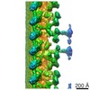
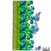
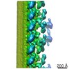
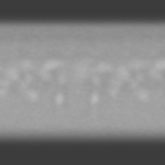
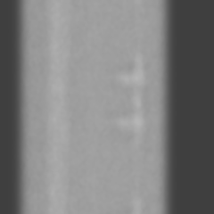
| Projections & slices | Image control
Images are generated by Spider. | ||||||||||||||||||||||||||||||||||||||||||||||||||||||||||||||||||||
| Voxel size | X=Y=Z: 6.07 Å | ||||||||||||||||||||||||||||||||||||||||||||||||||||||||||||||||||||
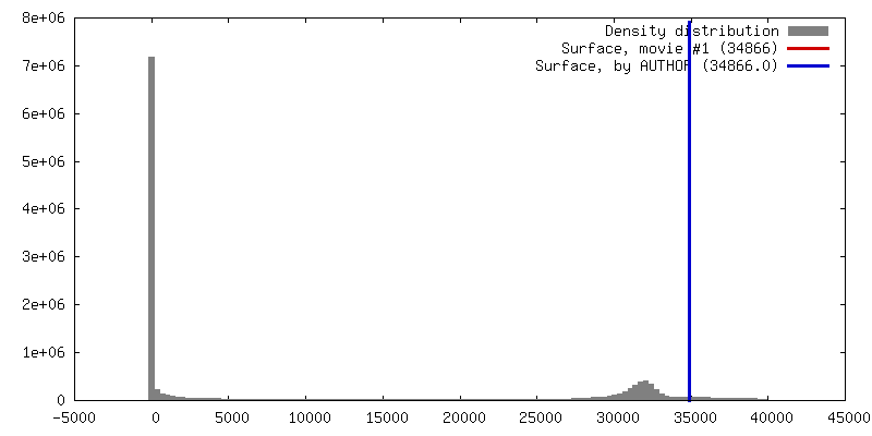
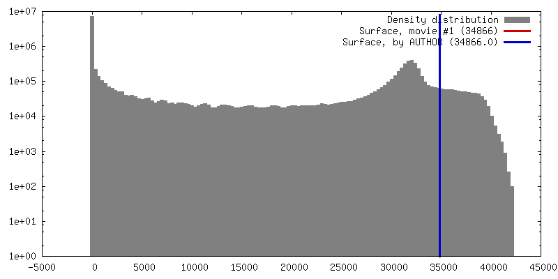
| Density |
| ||||||||||||||||||||||||||||||||||||||||||||||||||||||||||||||||||||
| Symmetry | Space group: 1 | ||||||||||||||||||||||||||||||||||||||||||||||||||||||||||||||||||||
| Details | EMDB XML:
CCP4 map header:
| ||||||||||||||||||||||||||||||||||||||||||||||||||||||||||||||||||||
 Z (Sec.)
 Y (Row.)
 X (Col.)


-Supplemental data
- Sample components
-Entire : Chlamydomonas axoneme, strain oda6-ic2-n-bccp, labeled with strep...
| Entire | Name: Chlamydomonas axoneme, strain oda6-ic2-n-bccp, labeled with streptavidin |
|---|---|
| Components |
|
-Supramolecule #1000: Chlamydomonas axoneme, strain oda6-ic2-n-bccp, labeled with strep...
| Supramolecule | Name: Chlamydomonas axoneme, strain oda6-ic2-n-bccp, labeled with streptavidin type: sample / ID: 1000 / Number unique components: 2 |
|---|
-Supramolecule #1: axoneme
| Supramolecule | Name: axoneme / type: organelle_or_cellular_component / ID: 1 / Recombinant expression: No / Database: NCBI |
|---|---|
| Source (natural) | Organism: Chlamydomonas reinhardtii (plant) / Strain: oda6-ic2-n-bccp / Organelle: flagella |
-Experimental details
-Structure determination
| Method | cryo EM |
|---|---|
 Processing Processing | subtomogram averaging |
| Aggregation state | filament |
-Sample preparation
| Concentration | 0.04 mg/mL |
|---|---|
| Buffer | pH: 7.2 Details: 30 mM Hepes-NaOH pH 7.2, 5 mM MgCl2, 1 mM dithiothreitol, 1 mM EGTA, 50 mM CH3COOK, and 1 mM phenylmethylsulfonyl fluoride |
| Grid | Details: 300 mesh copper grid with home-made holey carbon support |
| Vitrification | Cryogen name: ETHANE / Chamber humidity: 80 % / Chamber temperature: 93 K / Instrument: LEICA EM GP / Method: Blot for 5 seconds before plunging |
- Electron microscopy
| Microscope | JEOL 3100FFC |
|---|---|
| Temperature | Min: 90 K / Max: 95 K |
| Date | Feb 20, 2013 |
| Image recording | Category: CCD / Film or detector model: TVIPS TEMCAM-F416 (4k x 4k) / Average electron dose: 90 e/Å2 |
| Electron beam | Acceleration voltage: 300 kV / Electron source:  FIELD EMISSION GUN FIELD EMISSION GUN |
| Electron optics | Calibrated magnification: 25700 / Illumination mode: FLOOD BEAM / Imaging mode: BRIGHT FIELD / Nominal magnification: 15000 |
| Sample stage | Specimen holder model: GATAN LIQUID NITROGEN / Tilt series - Axis1 - Min angle: -60.5 ° / Tilt series - Axis1 - Max angle: 60.5 ° |
-Image processing
| Final reconstruction | Resolution.type: BY AUTHOR / Resolution: 57.0 Å / Resolution method: FSC 0.5 CUT-OFF / Software - Name: PEET, Ruby-Helix / Number subtomograms used: 77 |
|---|
