ムービー
ムービー コントローラー


+ データを開く

- 基本情報
基本情報
| 登録情報 | データベース: EMDB / ID: EMD-5699 | |||||||||
|---|---|---|---|---|---|---|---|---|---|---|
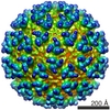
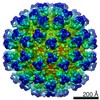
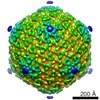
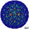
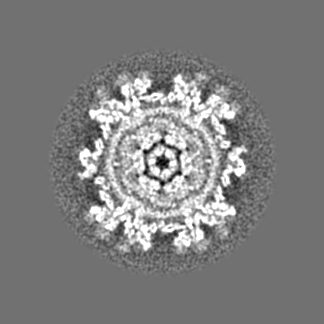
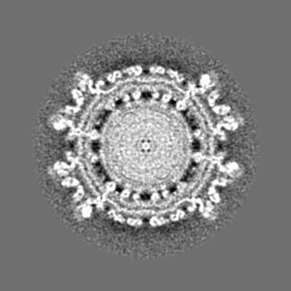
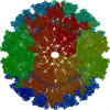
| タイトル | CryoEM structure of EGFR-specific virus (derived from sindbis virus) | |||||||||
 マップデータ マップデータ | CryoEM structure of EGFR-specific virus derived from sindbis virus | |||||||||
 試料 試料 |
| |||||||||
 キーワード キーワード | EGFR-specific virus / sindbis virus | |||||||||
| 生物種 | synthetic construct (人工物) | |||||||||
| 手法 | 単粒子再構成法 / クライオ電子顕微鏡法 / 解像度: 12.2 Å | |||||||||
 データ登録者 データ登録者 | Dai H / Liu Z / Jiang W / Kuhn RJ | |||||||||
 引用 引用 | ジャーナル: J Virol / 年: 2013 タイトル: Directed evolution of a virus exclusively utilizing human epidermal growth factor receptor as the entry receptor. 著者: Hong-Sheng Dai / Zheng Liu / Wen Jiang / Richard J Kuhn 要旨: Rational design and directed evolution are powerful tools to generate and improve protein function; however, their uses are mostly limited to enzyme and antibody engineering. Here we describe a ...Rational design and directed evolution are powerful tools to generate and improve protein function; however, their uses are mostly limited to enzyme and antibody engineering. Here we describe a directed-evolution strategy, named the tandem selection and enrichment system (TSES), and its use in generating virus with exclusive specificity for a particular cellular receptor. In TSES, evolving viruses are sequentially and iteratively transferred between two different host cells, one for selection of receptor specificity and the other for enrichment of the fittest virus. By combining rational design and TSES, we generated human epidermal growth factor receptor (EGFR)-specific virus 1 (ESV1). ESV1 has the backbone of Sindbis virus (SINV) and displays an EGF domain engrafted onto structural protein E2 after residue Pro192, together with eight amino acid changes stabilizing the E2-EGF chimera. ESV1 uses EGFR to initiate infection and has lost the capacity to interact with all known SINV receptors. A 12.2-Å cryoelectron microscopic (cryoEM) reconstruction of ESV1 reveals that the E2-EGF fusion adopts a fixed conformation, with EGF sitting at the top of the E2 spike; The EGFR binding interface faces outward, and the EGF domain completely masks SINV receptor binding. The cryoEM structure of ESV1 explains the desirable properties of ESV1 and provides insights for its further modification. TSES expands the scope of directed evolution and can be easily extended to other targeting molecules and viral systems. | |||||||||
| 履歴 |
|
- 構造の表示
構造の表示
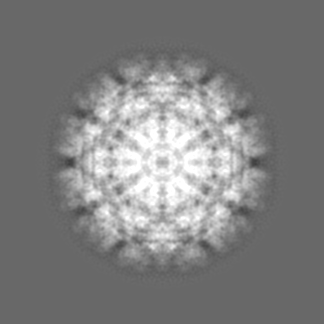
| ムービー |
 ムービービューア ムービービューア |
|---|---|
| 構造ビューア | EMマップ: SurfViewMolmilJmol/JSmol |
| 添付画像 |

- ダウンロードとリンク
-EMDBアーカイブ
| マップデータ | emd_5699.map.gz | 33.6 MB | EMDBマップデータ形式 | |
|---|---|---|---|---|
| ヘッダ (付随情報) | emd-5699-v30.xmlemd-5699.xml | 11.2 KB 11.2 KB | 表示 表示 | EMDBヘッダ |
| 画像 | 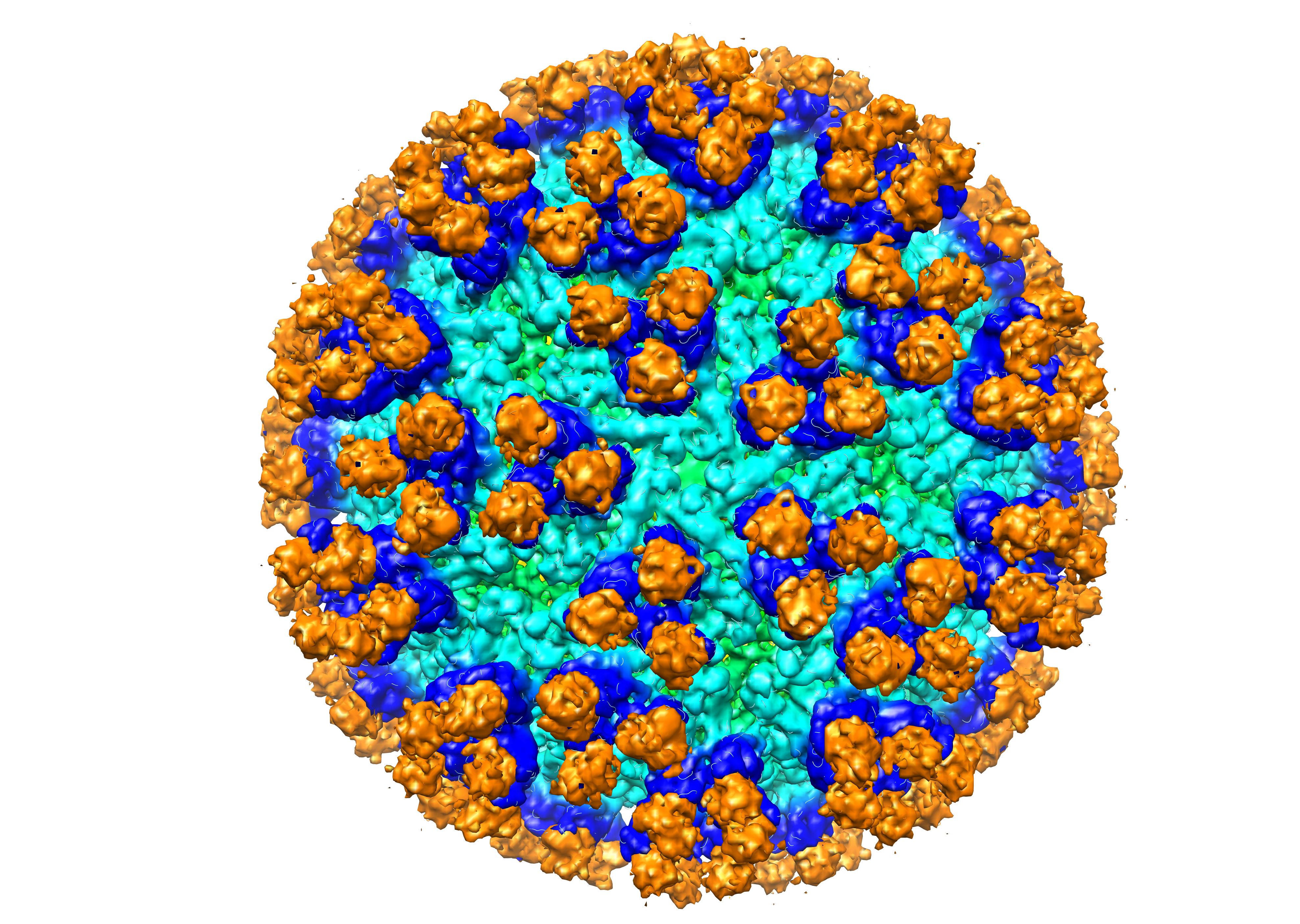 emd_5699.png emd_5699.png | 5.5 MB | ||
| アーカイブディレクトリ |  http://ftp.pdbj.org/pub/emdb/structures/EMD-5699ftp://ftp.pdbj.org/pub/emdb/structures/EMD-5699 http://ftp.pdbj.org/pub/emdb/structures/EMD-5699ftp://ftp.pdbj.org/pub/emdb/structures/EMD-5699 | HTTPS FTP |
-検証レポート
| 文書・要旨 | emd_5699_validation.pdf.gz | 77.9 KB | 表示 | EMDB検証レポート |
|---|---|---|---|---|
| 文書・詳細版 | emd_5699_full_validation.pdf.gz | 77 KB | 表示 | |
| XML形式データ | emd_5699_validation.xml.gz | 494 B | 表示 | |
| アーカイブディレクトリ | https://ftp.pdbj.org/pub/emdb/validation_reports/EMD-5699ftp://ftp.pdbj.org/pub/emdb/validation_reports/EMD-5699 | HTTPS FTP |
-関連構造データ

-リンク
| EMDBのページ | EMDB (EBI/PDBe) / EMDataResource |
|---|
-マップ
| ファイル | ダウンロード / ファイル: emd_5699.map.gz / 形式: CCP4 / 大きさ: 126.7 MB / タイプ: IMAGE STORED AS FLOATING POINT NUMBER (4 BYTES) | ||||||||||||||||||||||||||||||||||||||||||||||||||||||||||||||||||||
|---|---|---|---|---|---|---|---|---|---|---|---|---|---|---|---|---|---|---|---|---|---|---|---|---|---|---|---|---|---|---|---|---|---|---|---|---|---|---|---|---|---|---|---|---|---|---|---|---|---|---|---|---|---|---|---|---|---|---|---|---|---|---|---|---|---|---|---|---|---|
| 注釈 | CryoEM structure of EGFR-specific virus derived from sindbis virus | ||||||||||||||||||||||||||||||||||||||||||||||||||||||||||||||||||||
| 投影像・断面図 | 画像のコントロール
画像は Spider により作成 | ||||||||||||||||||||||||||||||||||||||||||||||||||||||||||||||||||||
| ボクセルのサイズ | X=Y=Z: 3.24 Å | ||||||||||||||||||||||||||||||||||||||||||||||||||||||||||||||||||||
| 密度 |
| ||||||||||||||||||||||||||||||||||||||||||||||||||||||||||||||||||||
| 対称性 | 空間群: 1 | ||||||||||||||||||||||||||||||||||||||||||||||||||||||||||||||||||||
| 詳細 | EMDB XML:
CCP4マップ ヘッダ情報:
| ||||||||||||||||||||||||||||||||||||||||||||||||||||||||||||||||||||
 Z (Sec.)
 Y (Row.)
 X (Col.)


-添付データ
- 試料の構成要素
-全体 : EGFR-specific virus (derived from sindbis virus)
| 全体 | 名称: EGFR-specific virus (derived from sindbis virus) |
|---|---|
| 要素 |
|
-超分子 #1000: EGFR-specific virus (derived from sindbis virus)
| 超分子 | 名称: EGFR-specific virus (derived from sindbis virus) / タイプ: sample / ID: 1000 / 詳細: The sample was monodisperse. / 集合状態: icosahedral / Number unique components: 1 |
|---|
-超分子 #1: synthetic construct
| 超分子 | 名称: synthetic construct / タイプ: virus / ID: 1 / Name.synonym: EGFR-specific Sindbis virus 詳細: EGF domain was engrafted onto an icosahedral virus scaffold, and directed evolution was conducted to stabilize the chimeric virus. NCBI-ID: 32630 / 生物種: synthetic construct / Sci species strain: EGFR-specific no.1 / データベース: NCBI / ウイルスタイプ: VIRION / ウイルス・単離状態: STRAIN / ウイルス・エンベロープ: Yes / ウイルス・中空状態: No / Syn species name: EGFR-specific Sindbis virus |
|---|---|
| 宿主 | 生物種:  Homo sapiens (ヒト) / 別称: VERTEBRATES Homo sapiens (ヒト) / 別称: VERTEBRATES |
| ウイルス殻 | Shell ID: 1 / 名称: E1 and E2-EGF / 直径: 375.00 Å / T番号(三角分割数): 4 |
-実験情報
-構造解析
| 手法 | クライオ電子顕微鏡法 |
|---|---|
 解析 解析 | 単粒子再構成法 |
| 試料の集合状態 | particle |
-試料調製
| 濃度 | 1.5 mg/mL |
|---|---|
| 緩衝液 | pH: 7.4 / 詳細: 50 mM Tris, 200 mM NaCl, 1 mM EDTA |
| グリッド | 詳細: 400 mesh copper grid with thin carbon support, glow discharged in air |
| 凍結 | 凍結剤: ETHANE / チャンバー内湿度: 70 % / チャンバー内温度: 97.15 K / 装置: GATAN CRYOPLUNGE 3 / 手法: Blot for 6 seconds before plunging. |
- 電子顕微鏡法
| 顕微鏡 | FEI/PHILIPS CM200FEG |
|---|---|
| 温度 | 最低: 93.15 K / 最高: 100.15 K / 平均: 97 K |
| アライメント法 | Legacy - 非点収差: Objective lens astigmatism was corrected at 100,000 times magnification |
| 日付 | 2011年10月12日 |
| 撮影 | カテゴリ: FILM / フィルム・検出器のモデル: KODAK SO-163 FILM デジタル化 - スキャナー: NIKON SUPER COOLSCAN 9000 デジタル化 - サンプリング間隔: 3.24 µm / 実像数: 166 / 平均電子線量: 20 e/Å2 / ビット/ピクセル: 16 |
| 電子線 | 加速電圧: 200 kV / 電子線源:  FIELD EMISSION GUN FIELD EMISSION GUN |
| 電子光学系 | 倍率(補正後): 39190 / 照射モード: FLOOD BEAM / 撮影モード: BRIGHT FIELD / Cs: 2.0 mm / 最大 デフォーカス(公称値): 5.0 µm / 最小 デフォーカス(公称値): 1.5 µm / 倍率(公称値): 38000 |
| 試料ステージ | 試料ホルダーモデル: SIDE ENTRY, EUCENTRIC |
-画像解析
| CTF補正 | 詳細: each particle |
|---|---|
| 最終 再構成 | アルゴリズム: OTHER / 解像度のタイプ: BY AUTHOR / 解像度: 12.2 Å / 解像度の算出法: OTHER / ソフトウェア - 名称: jspr, EMAN2, EMAN / 詳細: Final maps were calculated from 2290 particles. / 使用した粒子像数: 2290 |
-原子モデル構築 1
| 初期モデル | PDB ID: |
|---|---|
| ソフトウェア | 名称: veda |
| 精密化 | 空間: REAL / プロトコル: RIGID BODY FIT |
-原子モデル構築 2
| 初期モデル | PDB ID: |
|---|---|
| ソフトウェア | 名称: Chimera |
| 精密化 | 空間: REAL / プロトコル: RIGID BODY FIT |