ムービー
ムービー コントローラー


+ データを開く

- 基本情報
基本情報
| 登録情報 | データベース: EMDB / ID: EMD-5680 | |||||||||
|---|---|---|---|---|---|---|---|---|---|---|
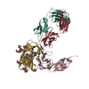
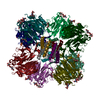
| タイトル | Cryo-electron microscopy of a trimeric soluble HIV Env construct, gp140 SOSIP, in complex with Fab Z13e1 | |||||||||
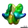
 マップデータ マップデータ | Reconstruction of trimeric HIV gp140 with MPER FAB | |||||||||
 試料 試料 |
| |||||||||
 キーワード キーワード | HIV / gp140 / SOSIP / Z13e1 | |||||||||
| 生物種 |   Human immunodeficiency virus 1 (ヒト免疫不全ウイルス) / unidentified (未定義) Human immunodeficiency virus 1 (ヒト免疫不全ウイルス) / unidentified (未定義) | |||||||||
| 手法 | 単粒子再構成法 / クライオ電子顕微鏡法 / 解像度: 18.5 Å | |||||||||
 データ登録者 データ登録者 | Harris AK / Bartesaghi A / Milne JL / Subramaniam S | |||||||||
 引用 引用 | ジャーナル: J Virol / 年: 2013 タイトル: HIV-1 envelope glycoprotein trimers display open quaternary conformation when bound to the gp41 membrane-proximal external-region-directed broadly neutralizing antibody Z13e1. 著者: Audray K Harris / Alberto Bartesaghi / Jacqueline L S Milne / Sriram Subramaniam /  要旨: We describe cryo-electron microscopic studies of the interaction between the ectodomain of the trimeric HIV-1 envelope glycoprotein (Env) and Z13e1, a broadly neutralizing antibody that targets the ...We describe cryo-electron microscopic studies of the interaction between the ectodomain of the trimeric HIV-1 envelope glycoprotein (Env) and Z13e1, a broadly neutralizing antibody that targets the membrane-proximal external region (MPER) of the gp41 subunit. We show that Z13e1-bound Env displays an open quaternary conformation similar to the CD4-bound conformation. Our results support the idea that MPER-directed antibodies, such as Z13e1, block viral entry by interacting with Env at a step after CD4 activation. | |||||||||
| 履歴 |
|
- 構造の表示
構造の表示
| ムービー |
 ムービービューア ムービービューア |
|---|---|
| 構造ビューア | EMマップ: SurfViewMolmilJmol/JSmol |
| 添付画像 |

- ダウンロードとリンク
-EMDBアーカイブ
| マップデータ | emd_5680.map.gz | 5.2 MB | EMDBマップデータ形式 | |
|---|---|---|---|---|
| ヘッダ (付随情報) | emd-5680-v30.xmlemd-5680.xml | 9.9 KB 9.9 KB | 表示 表示 | EMDBヘッダ |
| 画像 | 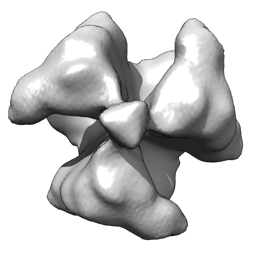 emd_5680_1.jpg emd_5680_1.jpg | 82 KB | ||
| アーカイブディレクトリ |  http://ftp.pdbj.org/pub/emdb/structures/EMD-5680ftp://ftp.pdbj.org/pub/emdb/structures/EMD-5680 http://ftp.pdbj.org/pub/emdb/structures/EMD-5680ftp://ftp.pdbj.org/pub/emdb/structures/EMD-5680 | HTTPS FTP |
-検証レポート
| 文書・要旨 | emd_5680_validation.pdf.gz | 78.7 KB | 表示 | EMDB検証レポート |
|---|---|---|---|---|
| 文書・詳細版 | emd_5680_full_validation.pdf.gz | 77.8 KB | 表示 | |
| XML形式データ | emd_5680_validation.xml.gz | 494 B | 表示 | |
| アーカイブディレクトリ | https://ftp.pdbj.org/pub/emdb/validation_reports/EMD-5680ftp://ftp.pdbj.org/pub/emdb/validation_reports/EMD-5680 | HTTPS FTP |
-関連構造データ


-リンク
| EMDBのページ | EMDB (EBI/PDBe) / EMDataResource |
|---|
-マップ
| ファイル | ダウンロード / ファイル: emd_5680.map.gz / 形式: CCP4 / 大きさ: 23.2 MB / タイプ: IMAGE STORED AS FLOATING POINT NUMBER (4 BYTES) | ||||||||||||||||||||||||||||||||||||||||||||||||||||||||||||||||||||
|---|---|---|---|---|---|---|---|---|---|---|---|---|---|---|---|---|---|---|---|---|---|---|---|---|---|---|---|---|---|---|---|---|---|---|---|---|---|---|---|---|---|---|---|---|---|---|---|---|---|---|---|---|---|---|---|---|---|---|---|---|---|---|---|---|---|---|---|---|---|
| 注釈 | Reconstruction of trimeric HIV gp140 with MPER FAB | ||||||||||||||||||||||||||||||||||||||||||||||||||||||||||||||||||||
| 投影像・断面図 | 画像のコントロール
画像は Spider により作成 | ||||||||||||||||||||||||||||||||||||||||||||||||||||||||||||||||||||
| ボクセルのサイズ | X=Y=Z: 1.35 Å | ||||||||||||||||||||||||||||||||||||||||||||||||||||||||||||||||||||
| 密度 |
| ||||||||||||||||||||||||||||||||||||||||||||||||||||||||||||||||||||
| 対称性 | 空間群: 1 | ||||||||||||||||||||||||||||||||||||||||||||||||||||||||||||||||||||
| 詳細 | EMDB XML:
CCP4マップ ヘッダ情報:
| ||||||||||||||||||||||||||||||||||||||||||||||||||||||||||||||||||||
 Z (Sec.)
 Y (Row.)
 X (Col.)


-添付データ
- 試料の構成要素
-全体 : Molecular structure of KNH1144 SOSIP gp140 with Z13e1 Fab
| 全体 | 名称: Molecular structure of KNH1144 SOSIP gp140 with Z13e1 Fab |
|---|---|
| 要素 |
|
-超分子 #1000: Molecular structure of KNH1144 SOSIP gp140 with Z13e1 Fab
| 超分子 | 名称: Molecular structure of KNH1144 SOSIP gp140 with Z13e1 Fab タイプ: sample / ID: 1000 / 集合状態: trimer / Number unique components: 2 |
|---|
-分子 #1: Envelope glycoprotein
| 分子 | 名称: Envelope glycoprotein / タイプ: protein_or_peptide / ID: 1 / Name.synonym: Env / コピー数: 3 / 集合状態: trimer / 組換発現: Yes |
|---|---|
| 由来(天然) | 生物種: Human immunodeficiency virus 1 (ヒト免疫不全ウイルス) 株: isolate 00KE_KNH1144 / 別称: HIV-1 |
| 分子量 | 実験値: 420 KDa |
| 組換発現 | 生物種:  Homo sapiens (ヒト) / 組換プラスミド: SOSIP-PPI4 and furin-pcDNA3.1 Homo sapiens (ヒト) / 組換プラスミド: SOSIP-PPI4 and furin-pcDNA3.1 |
-分子 #2: Fab portion of monoclonal antibody Z13e1
| 分子 | 名称: Fab portion of monoclonal antibody Z13e1 / タイプ: protein_or_peptide / ID: 2 / Name.synonym: Z13e1 Fab / 詳細: Fab fragment / 組換発現: No / データベース: NCBI |
|---|---|
| 由来(天然) | 生物種: unidentified (未定義) |
| 分子量 | 理論値: 50 KDa |
-実験情報
-構造解析
| 手法 | クライオ電子顕微鏡法 |
|---|---|
 解析 解析 | 単粒子再構成法 |
| 試料の集合状態 | particle |
-試料調製
| 濃度 | 0.42 mg/mL |
|---|---|
| 緩衝液 | pH: 7.5 / 詳細: TNE Buffer (10 mM Tris, 150 mM NaCl, 1 mM EDTA) |
| グリッド | 詳細: Quantifoil, plasma cleaned |
| 凍結 | 凍結剤: ETHANE / チャンバー内湿度: 100 % / 装置: FEI VITROBOT MARK III 手法: Blot for 6 seconds (blot offset -2) and plunge into an ethane slurry cooled by liquid nitrogen. |
- 電子顕微鏡法
| 顕微鏡 | FEI TITAN KRIOS |
|---|---|
| 日付 | 2009年8月31日 |
| 撮影 | カテゴリ: CCD フィルム・検出器のモデル: GATAN ULTRASCAN 4000 (4k x 4k) 実像数: 583 / 平均電子線量: 20 e/Å2 |
| 電子線 | 加速電圧: 80 kV / 電子線源:  FIELD EMISSION GUN FIELD EMISSION GUN |
| 電子光学系 | 照射モード: FLOOD BEAM / 撮影モード: BRIGHT FIELD / 最大 デフォーカス(公称値): 2.9 µm / 最小 デフォーカス(公称値): 1.3 µm / 倍率(公称値): 59500 |
| 試料ステージ | 試料ホルダーモデル: FEI TITAN KRIOS AUTOGRID HOLDER |
| 実験機器 |  モデル: Titan Krios / 画像提供: FEI Company |
-画像解析
| 最終 再構成 | 解像度のタイプ: BY AUTHOR / 解像度: 18.5 Å / 解像度の算出法: FSC 0.5 CUT-OFF / ソフトウェア - 名称: EMAN1 / 使用した粒子像数: 7050 |
|---|
-原子モデル構築 1
| 初期モデル | PDB ID: |
|---|---|
| ソフトウェア | 名称: Chimera |
| 詳細 | Protocol: Rigid body. Automated fitting procedures |
| 精密化 | 空間: REAL / プロトコル: RIGID BODY FIT |