 Movie
Movie Controller


+ Open data

- Basic information
| Entry | Database: EMDB / ID: EMD-5650 | |||||||||
|---|---|---|---|---|---|---|---|---|---|---|
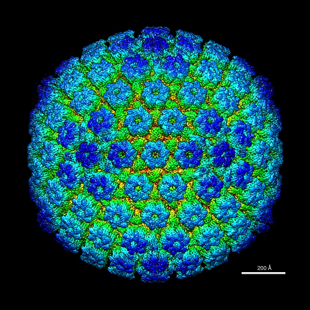
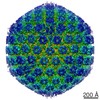
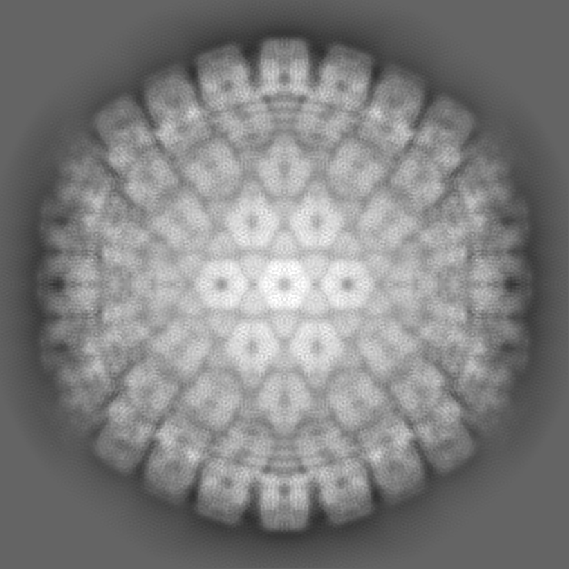
| Title | Pseudorabies virus C-capsid from cryo-electron microscopy | |||||||||
 Map data Map data | Reconstruction of pseudorabies virus (PRV) C-capsid | |||||||||
 Sample Sample |
| |||||||||
 Keywords Keywords | herpesvirus / capsid | |||||||||
| Biological species |  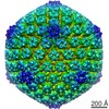 Suid herpesvirus 1 Suid herpesvirus 1 | |||||||||
| Method | single particle reconstruction / cryo EM / Resolution: 9.0 Å | |||||||||
 Authors Authors | Homa FL / Huffman JB / Toropova K / Lopez HR / Makhov AM / Conway JF | |||||||||
 Citation Citation | Journal: J Mol Biol / Year: 2013 Title: Structure of the pseudorabies virus capsid: comparison with herpes simplex virus type 1 and differential binding of essential minor proteins. Authors: F L Homa / J B Huffman / K Toropova / H R Lopez / A M Makhov / J F Conway /  Abstract: The structure of pseudorabies virus (PRV) capsids isolated from the nucleus of infected cells and from PRV virions was determined by cryo-electron microscopy (cryo-EM) and compared to herpes simplex ...The structure of pseudorabies virus (PRV) capsids isolated from the nucleus of infected cells and from PRV virions was determined by cryo-electron microscopy (cryo-EM) and compared to herpes simplex virus type 1 (HSV-1) capsids. PRV capsid structures closely resemble those of HSV-1, including distribution of the capsid vertex specific component (CVSC) of HSV-1, which is a heterodimer of the pUL17 and pUL25 proteins. Occupancy of CVSC on all PRV capsids is near 100%, compared to ~50% reported for HSV-1 C-capsids and 25% or less that we measure for HSV-1 A- and B-capsids. A PRV mutant lacking pUL25 does not produce C-capsids and lacks visible CVSC density in the cryo-EM-based reconstruction. A reconstruction of PRV capsids in which green fluorescent protein was fused within the N-terminus of pUL25 confirmed previous studies with a similar HSV-1 capsid mutant localizing pUL25 to the CVSC density region that is distal to the penton. However, comparison of the CVSC density in a 9-Å-resolution PRV C-capsid map with the available crystal structure of HSV-1 pUL25 failed to find a satisfactory fit, suggesting either a different fold for PRV pUL25 or a capsid-bound conformation for pUL25 that does not match the X-ray model determined from protein crystallized in solution. The PRV capsid imaged within virions closely resembles C-capsids with the addition of weak but significant density shrouding the pentons that we attribute to tegument proteins. Our results demonstrate significant structure conservation between the PRV and HSV capsids. | |||||||||
| History |
|
- Structure visualization
| Movie |
 Movie viewer Movie viewer |
|---|---|
| Structure viewer | EM map: SurfViewMolmilJmol/JSmol |
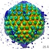
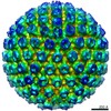
| Supplemental images |
- Downloads & links
-EMDB archive
| Map data | emd_5650.map.gz | 634.4 MB | EMDB map data format | |
|---|---|---|---|---|
| Header (meta data) | emd-5650-v30.xmlemd-5650.xml | 9.2 KB 9.2 KB | Display Display | EMDB header |
| Images | emd_5650.tif | 1.7 MB | ||
| Archive directory |  http://ftp.pdbj.org/pub/emdb/structures/EMD-5650ftp://ftp.pdbj.org/pub/emdb/structures/EMD-5650 http://ftp.pdbj.org/pub/emdb/structures/EMD-5650ftp://ftp.pdbj.org/pub/emdb/structures/EMD-5650 | HTTPS FTP |
-Validation report
| Summary document | emd_5650_validation.pdf.gz | 78.2 KB | Display | EMDB validaton report |
|---|---|---|---|---|
| Full document | emd_5650_full_validation.pdf.gz | 77.3 KB | Display | |
| Data in XML | emd_5650_validation.xml.gz | 493 B | Display | |
| Arichive directory | https://ftp.pdbj.org/pub/emdb/validation_reports/EMD-5650ftp://ftp.pdbj.org/pub/emdb/validation_reports/EMD-5650 | HTTPS FTP |
-Related structure data
| Related structure data | 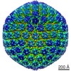 5652C 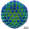 5654C 5655C 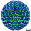 5656C 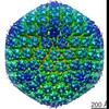 5657C 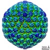 5659C 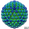 5660C 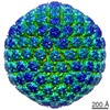 5661C C: citing same article ( |
|---|---|
| Similar structure data |

-Links
| EMDB pages | EMDB (EBI/PDBe) / EMDataResource |
|---|
-Map
| File | Download / File: emd_5650.map.gz / Format: CCP4 / Size: 1 GB / Type: IMAGE STORED AS SIGNED INTEGER (2 BYTES) | ||||||||||||||||||||||||||||||||||||||||||||||||||||||||||||||||||||
|---|---|---|---|---|---|---|---|---|---|---|---|---|---|---|---|---|---|---|---|---|---|---|---|---|---|---|---|---|---|---|---|---|---|---|---|---|---|---|---|---|---|---|---|---|---|---|---|---|---|---|---|---|---|---|---|---|---|---|---|---|---|---|---|---|---|---|---|---|---|
| Annotation | Reconstruction of pseudorabies virus (PRV) C-capsid | ||||||||||||||||||||||||||||||||||||||||||||||||||||||||||||||||||||
| Projections & slices | Image control
Images are generated by Spider. | ||||||||||||||||||||||||||||||||||||||||||||||||||||||||||||||||||||
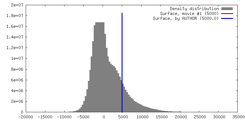
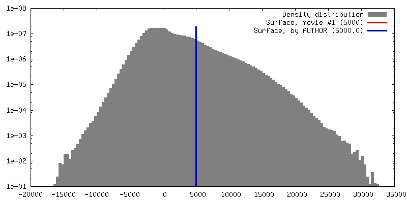
| Voxel size | X=Y=Z: 1.63 Å | ||||||||||||||||||||||||||||||||||||||||||||||||||||||||||||||||||||
| Density |
| ||||||||||||||||||||||||||||||||||||||||||||||||||||||||||||||||||||
| Symmetry | Space group: 1 | ||||||||||||||||||||||||||||||||||||||||||||||||||||||||||||||||||||
| Details | EMDB XML:
CCP4 map header:
| ||||||||||||||||||||||||||||||||||||||||||||||||||||||||||||||||||||
 Z (Sec.)
 Y (Row.)
 X (Col.)


-Supplemental data
- Sample components
-Entire : Pseudorabies virus (PRV) C-capsid
| Entire | Name: Pseudorabies virus (PRV) C-capsid |
|---|---|
| Components |
|
-Supramolecule #1000: Pseudorabies virus (PRV) C-capsid
| Supramolecule | Name: Pseudorabies virus (PRV) C-capsid / type: sample / ID: 1000 / Number unique components: 1 |
|---|
-Supramolecule #1: Suid herpesvirus 1
| Supramolecule | Name: Suid herpesvirus 1 / type: virus / ID: 1 / Name.synonym: pseudorabies virus / Details: C-capsid contains viral dsDNA. / NCBI-ID: 10345 / Sci species name: Suid herpesvirus 1 / Sci species strain: Becker / Database: NCBI / Virus type: VIRION / Virus isolate: STRAIN / Virus enveloped: Yes / Virus empty: No / Syn species name: pseudorabies virus |
|---|---|
| Host (natural) | Organism:  |
| Virus shell | Shell ID: 1 / Name: C-capsid / Diameter: 1250 Å / T number (triangulation number): 16 |
-Experimental details
-Structure determination
| Method | cryo EM |
|---|---|
 Processing Processing | single particle reconstruction |
| Aggregation state | particle |
-Sample preparation
| Buffer | pH: 7.5 / Details: TNE: 500mM NaCl, 10mM Tris, 1mM EDTA |
|---|---|
| Grid | Details: Quantifoil R2/1, 200 mesh copper, glow discharged for 10-15 secs |
| Vitrification | Cryogen name: ETHANE-PROPANE MIXTURE / Chamber humidity: 95 % / Chamber temperature: 82 K / Instrument: FEI VITROBOT MARK III / Method: Blot for 6 seconds. |
- Electron microscopy
| Microscope | FEI POLARA 300 |
|---|---|
| Date | Jul 18, 2010 |
| Image recording | Category: FILM / Film or detector model: KODAK SO-163 FILM / Digitization - Scanner: NIKON SUPER COOLSCAN 9000 / Digitization - Sampling interval: 6.35 µm / Number real images: 535 / Bits/pixel: 8 |
| Electron beam | Acceleration voltage: 300 kV / Electron source:  FIELD EMISSION GUN FIELD EMISSION GUN |
| Electron optics | Illumination mode: FLOOD BEAM / Imaging mode: BRIGHT FIELD / Cs: 2 mm / Nominal magnification: 39000 |
| Sample stage | Specimen holder: Polara stage / Specimen holder model: GATAN LIQUID NITROGEN |
| Experimental equipment |  Model: Tecnai Polara / Image courtesy: FEI Company |
-Image processing
| Details | Particles were selected manually with x3dpreprocess. |
|---|---|
| CTF correction | Details: Each micrograph |
| Final reconstruction | Algorithm: OTHER / Resolution.type: BY AUTHOR / Resolution: 9.0 Å / Resolution method: FSC 0.5 CUT-OFF / Software - Name: AUTO3DEM / Number images used: 11908 |
