H2020 Marie Curie Actions of the European Commission
101153233
European Union
Citation
Journal: Proc Natl Acad Sci U S A / Year: 2026 Title: SUN5 forms a regular protein lattice reinforcing the sperm head-tail junction. Authors: Jonas Moecking / Svetlana Doroshev / Miguel Ricardo Leung / Tzviya Zeev-Ben-Mordehai / Abstract: Spermatozoa strongly rely on their streamlined morphology to successfully fertilize an oocyte. A striking example of a morphological defect resulting in infertility is acephalic spermatozoa syndrome, ...Spermatozoa strongly rely on their streamlined morphology to successfully fertilize an oocyte. A striking example of a morphological defect resulting in infertility is acephalic spermatozoa syndrome, a rare but severe condition leading to detachment of the sperm head and tail. Among the most common genetic causes for this syndrome are mutations in the linker of nucleo- and cytoskeleton (LINC) complex component SUN5. LINC complexes typically reside in the nuclear envelope, the double-membrane surrounding the nucleus, where they establish a physical bridge between nucleus and cytoplasm. This localization allows them to transduce mechanical signals from the cytoplasm to the nucleus and to regulate nuclear morphology. In sperm, LINC complexes are essential for a multitude of morphological changes during sperm development, including the reshaping of the nucleus and establishing a stable head-tail junction. Here, using superresolution fluorescence microscopy, we find that sperm-specific SUN5 localizes to the base of the head in human, mouse, and boar sperm. By applying in situ cryoelectron tomography, we find an extensive hexagonal lattice in the nuclear envelope in this region. This lattice appears to maintain a consistent close apposition between the inner and outer nuclear membranes (ONM). Further structural analysis supports a model in which LINC complexes form this lattice by laterally interacting at the ONM. Overall, this study sheds light on nuclear envelope organization in the highly streamlined sperm cell, providing a structural basis for uniform nuclear envelope spacing maintained by a LINC lattice and rationalizing the disruptive effects of SUN5 mutations.
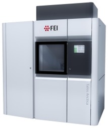
Model: Talos Arctica / Image courtesy: FEI Company
+
Image processing
Final reconstruction
Applied symmetry - Point group: C3 (3 fold cyclic) / Resolution.type: BY AUTHOR / Resolution: 13.7 Å / Resolution method: FSC 0.143 CUT-OFF / Software - Name: RELION (ver. 5.0.0) / Number subtomograms used: 15488
Extraction
Number tomograms: 44 / Number images used: 28361 / Method: Surface model / Software - Name: RELION (ver. 5.0.0) Details: The outer nuclear membrane was defined as a surface and particles were seeded using Dynamo. Particle coordinates were then transferred to Relion 5.0
In the structure databanks used in Yorodumi, some data are registered as the other names, "COVID-19 virus" and "2019-nCoV". Here are the details of the virus and the list of structure data.
Jan 31, 2019. EMDB accession codes are about to change! (news from PDBe EMDB page)
EMDB accession codes are about to change! (news from PDBe EMDB page)
The allocation of 4 digits for EMDB accession codes will soon come to an end. Whilst these codes will remain in use, new EMDB accession codes will include an additional digit and will expand incrementally as the available range of codes is exhausted. The current 4-digit format prefixed with “EMD-” (i.e. EMD-XXXX) will advance to a 5-digit format (i.e. EMD-XXXXX), and so on. It is currently estimated that the 4-digit codes will be depleted around Spring 2019, at which point the 5-digit format will come into force.
The EM Navigator/Yorodumi systems omit the EMD- prefix.
Related info.:Q: What is EMD? / ID/Accession-code notation in Yorodumi/EM Navigator
Yorodumi is a browser for structure data from EMDB, PDB, SASBDB, etc.
This page is also the successor to EM Navigator detail page, and also detail information page/front-end page for Omokage search.
The word "yorodu" (or yorozu) is an old Japanese word meaning "ten thousand". "mi" (miru) is to see.
Related info.:EMDB / PDB / SASBDB / Comparison of 3 databanks / Yorodumi Search / Aug 31, 2016. New EM Navigator & Yorodumi / Yorodumi Papers / Jmol/JSmol / Function and homology information / Changes in new EM Navigator and Yorodumi
 Movie
Movie Controller

 Yorodumi
 Open data
 Basic information
 Map data
 Sample
 Keywords
 Homo sapiens (human)
 Authors
 Citation
 Structure visualization
 Downloads & links
 EMDB map data format
 emd_56104.png
 https://data.pdbj.org/pub/emdb/structures/EMD-56104
 Z (Sec.)
 Y (Row.)
 X (Col.)


 Sample components
 Processing
 Electron microscopy
 FIELD EMISSION GUN