Movie
 Controller


+ Open data

- Basic information
| Entry |  | |||||||||
|---|---|---|---|---|---|---|---|---|---|---|
| Title | cryo-EM structure of human Tryptophan 2,3-dioxygenase (TDO2) | |||||||||
 Map data | ||||||||||
 Sample |
| |||||||||
 Keywords | Tryptophan metabolism / Heme cofactor / OXIDOREDUCTASE | |||||||||
| Function / homology |  Function and homology information response to nitroglycerin / tryptophan 2,3-dioxygenase / response to cortisol / L-tryptophan 2,3-dioxygenase activity / L-tryptophan catabolic process / Tryptophan catabolism / amino acid binding / response to dexamethasone / protein homotetramerization / response to ethanol ...response to nitroglycerin / tryptophan 2,3-dioxygenase / response to cortisol / L-tryptophan 2,3-dioxygenase activity / L-tryptophan catabolic process / Tryptophan catabolism / amino acid binding / response to dexamethasone / protein homotetramerization / response to ethanol / heme binding / metal ion binding / identical protein binding / cytosol Similarity search - Function | |||||||||
| Biological species |  Homo sapiens (human) | |||||||||
| Method | single particle reconstruction / cryo EM / Resolution: 3.25 Å | |||||||||
 Authors | Thielen AS / Prabu JR / Schulman BA | |||||||||
| Funding support |  Germany, 2 items
| |||||||||
 Citation | Journal: Mol Cell / Year: 2026 Title: A CK2-β-TrCP kinase-E3 ubiquitin ligase cascade is a metabolic sensor regulating tryptophan 2,3-dioxygenase stability. Authors: Alina S Thielen / Bastian Bräuning / Karthik V Gottemukkala / Judith Müller / J Rajan Prabu / Pooja Verma / Simon Klaessens / Benoit J Van den Eynde / Gary Kleiger / Peter J Murray / Brenda A Schulman /   Abstract: Small molecules toggling the ubiquitin-proteasome system (UPS) are powerful regulators of protein degradation, yet how endogenous ligands gate UPS decisions remains poorly understood. Here, we define ...Small molecules toggling the ubiquitin-proteasome system (UPS) are powerful regulators of protein degradation, yet how endogenous ligands gate UPS decisions remains poorly understood. Here, we define control of UPS access to human tryptophan 2,3-dioxygenase (TDO2), which converts the amino acid tryptophan (Trp) to N-formylkynurenine. When Trp concentrations are limiting, TDO2 is degraded to avert hypotryptophanemia. We discovered that a CK2-β-TrCP kinase-E3 ligase cascade generates and recognizes phosphodegrons in TDO2 unless they are shielded by Trp binding at an exosite distal from the catalytic site. Effects of Trp analogs on CK2-β-TrCP-dependent ubiquitylation indicated that the indole, amino, and carboxylate groups collectively protect TDO2. Cryo-EM reveals that these moieties organize a Trp-ordered flap that covers the exosite and thereby restricts generation of adjacent phosphodegrons. In the absence of Trp, the flap is flexible, enabling phosphorylation-coupled ubiquitylation. Overall, our data uncovered an endogenous small molecule conformationally stabilizing its own metabolizing enzyme through protection from a phosphorylation-ubiquitylation cascade. | |||||||||
| History |
|
- Structure visualization
| Supplemental images |
|---|

- Downloads & links
-EMDB archive
| Map data | emd_56042.map.gz | 32.8 MB | EMDB map data format | |
|---|---|---|---|---|
| Header (meta data) | emd-56042-v30.xmlemd-56042.xml | 20.6 KB 20.6 KB | Display Display | EMDB header |
| FSC (resolution estimation) | emd_56042_fsc.xml | 9.6 KB | Display | FSC data file |
| Images |  emd_56042.png | 70.4 KB | ||
| Masks | emd_56042_msk_1.map | 64 MB | Mask map | |
| Filedesc metadata | emd-56042.cif.gz | 6 KB | ||
| Others | emd_56042_additional_1.map.gzemd_56042_half_map_1.map.gzemd_56042_half_map_2.map.gz | 53.9 MB 59.4 MB 59.4 MB | ||
| Archive directory |  https://data.pdbj.org/pub/emdb/structures/EMD-56042ftp://data.pdbj.org/pub/emdb/structures/EMD-56042 | HTTPS FTP |
-Related structure data
| Related structure data |  9tl5MC  9tlfC  9tn8C C: citing same article ( M: atomic model generated by this map |
|---|---|
| Similar structure data |


-Links
| EMDB pages | EMDB (EBI/PDBe) / EMDataResource |
|---|
-Map
| File | Download / File: emd_56042.map.gz / Format: CCP4 / Size: 64 MB / Type: IMAGE STORED AS FLOATING POINT NUMBER (4 BYTES) | ||||||||||||||||||||||||||||||||||||
|---|---|---|---|---|---|---|---|---|---|---|---|---|---|---|---|---|---|---|---|---|---|---|---|---|---|---|---|---|---|---|---|---|---|---|---|---|---|
| Projections & slices | Image control
Images are generated by Spider. | ||||||||||||||||||||||||||||||||||||
| Voxel size | X=Y=Z: 0.8222 Å | ||||||||||||||||||||||||||||||||||||
| Density |
| ||||||||||||||||||||||||||||||||||||
| Symmetry | Space group: 1 | ||||||||||||||||||||||||||||||||||||
| Details | EMDB XML:
|
 Z (Sec.)
 Y (Row.)
 X (Col.)


-Supplemental data
-Mask #1
| File | emd_56042_msk_1.map | ||||||||||||
|---|---|---|---|---|---|---|---|---|---|---|---|---|---|
| Projections & Slices |
| ||||||||||||
| Density Histograms |


-Additional map: #1
| File | emd_56042_additional_1.map | ||||||||||||
|---|---|---|---|---|---|---|---|---|---|---|---|---|---|
| Projections & Slices |
| ||||||||||||
| Density Histograms |


-Half map: #1
| File | emd_56042_half_map_1.map | ||||||||||||
|---|---|---|---|---|---|---|---|---|---|---|---|---|---|
| Projections & Slices |
| ||||||||||||
| Density Histograms |


-Half map: #2
| File | emd_56042_half_map_2.map | ||||||||||||
|---|---|---|---|---|---|---|---|---|---|---|---|---|---|
| Projections & Slices |
| ||||||||||||
| Density Histograms |


- Sample components
-Entire : Tetrameric Tryptophan 2,3-dioxygenase
| Entire | Name: Tetrameric Tryptophan 2,3-dioxygenase |
|---|---|
| Components |
|
-Supramolecule #1: Tetrameric Tryptophan 2,3-dioxygenase
| Supramolecule | Name: Tetrameric Tryptophan 2,3-dioxygenase / type: complex / ID: 1 / Parent: 0 / Macromolecule list: #1 |
|---|---|
| Source (natural) | Organism: Homo sapiens (human) |
-Macromolecule #1: Tryptophan 2,3-dioxygenase
| Macromolecule | Name: Tryptophan 2,3-dioxygenase / type: protein_or_peptide / ID: 1 / Number of copies: 4 / Enantiomer: LEVO / EC number: tryptophan 2,3-dioxygenase |
|---|---|
| Source (natural) | Organism: Homo sapiens (human) |
| Molecular weight | Theoretical: 49.707316 KDa |
| Recombinant expression | Organism:   Escherichia coli (E. coli) |
| Sequence | String: HHHHHHENLY FQGSMSGCPF LGNNFGYTFK KLPVEGSEED KSQTGVNRAS KGGLIYGNYL HLEKVLNAQE LQSETKGNKI HDEHLFIIT HQAYELWFKQ ILWELDSVRE IFQNGHVRDE RNMLKVVSRM HRVSVILKLL VQQFSILETM TALDFNDFRE Y LSPASGFQ ...String: HHHHHHENLY FQGSMSGCPF LGNNFGYTFK KLPVEGSEED KSQTGVNRAS KGGLIYGNYL HLEKVLNAQE LQSETKGNKI HDEHLFIIT HQAYELWFKQ ILWELDSVRE IFQNGHVRDE RNMLKVVSRM HRVSVILKLL VQQFSILETM TALDFNDFRE Y LSPASGFQ SLQFRLLENK IGVLQNMRVP YNRRHYRDNF KGEENELLLK SEQEKTLLEL VEAWLERTPG LEPHGFNFWG KL EKNITRG LEEEFIRIQA KEESEEKEEQ VAEFQKQKEV LLSLFDEKRH EHLLSKGERR LSYRALQGAL MIYFYREEPR FQV PFQLLT SLMDIDSLMT KWRYNHVCMV HRMLGSKAGT GGSSGYHYLR STVSDRYKVF VDLFNLSTYL IPRHWIPKMN PTIH KFLYT AEYCDSSYFS SDESD UniProtKB: Tryptophan 2,3-dioxygenase |
-Macromolecule #2: alpha-methyl-L-tryptophan
| Macromolecule | Name: alpha-methyl-L-tryptophan / type: ligand / ID: 2 / Number of copies: 4 / Formula: ZIQ |
|---|---|
| Molecular weight | Theoretical: 218.252 Da |
| Chemical component information |  ChemComp-ZIQ: |
-Macromolecule #3: PROTOPORPHYRIN IX CONTAINING FE
| Macromolecule | Name: PROTOPORPHYRIN IX CONTAINING FE / type: ligand / ID: 3 / Number of copies: 4 / Formula: HEM |
|---|---|
| Molecular weight | Theoretical: 616.487 Da |
| Chemical component information | 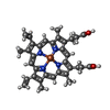 ChemComp-HEM: |
-Experimental details
-Structure determination
| Method | cryo EM |
|---|---|
 Processing | single particle reconstruction |
| Aggregation state | particle |
-Sample preparation
| Buffer | pH: 7.5 |
|---|---|
| Vitrification | Cryogen name: ETHANE-PROPANE |
- Electron microscopy
| Microscope | TFS KRIOS |
|---|---|
| Image recording | Film or detector model: GATAN K3 BIOQUANTUM (6k x 4k) / Average electron dose: 59.93 e/Å2 |
| Electron beam | Acceleration voltage: 300 kV / Electron source:  FIELD EMISSION GUN |
| Electron optics | Illumination mode: FLOOD BEAM / Imaging mode: BRIGHT FIELD / Nominal defocus max: 2.2 µm / Nominal defocus min: 0.6 µm |
| Experimental equipment | 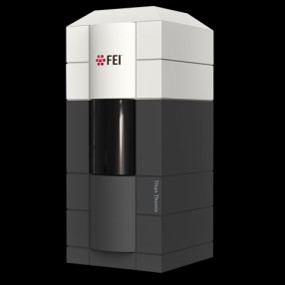 Model: Titan Krios / Image courtesy: FEI Company |