 ムービー
ムービー コントローラー


+ データを開く

- 基本情報
基本情報
| 登録情報 |  | |||||||||
|---|---|---|---|---|---|---|---|---|---|---|
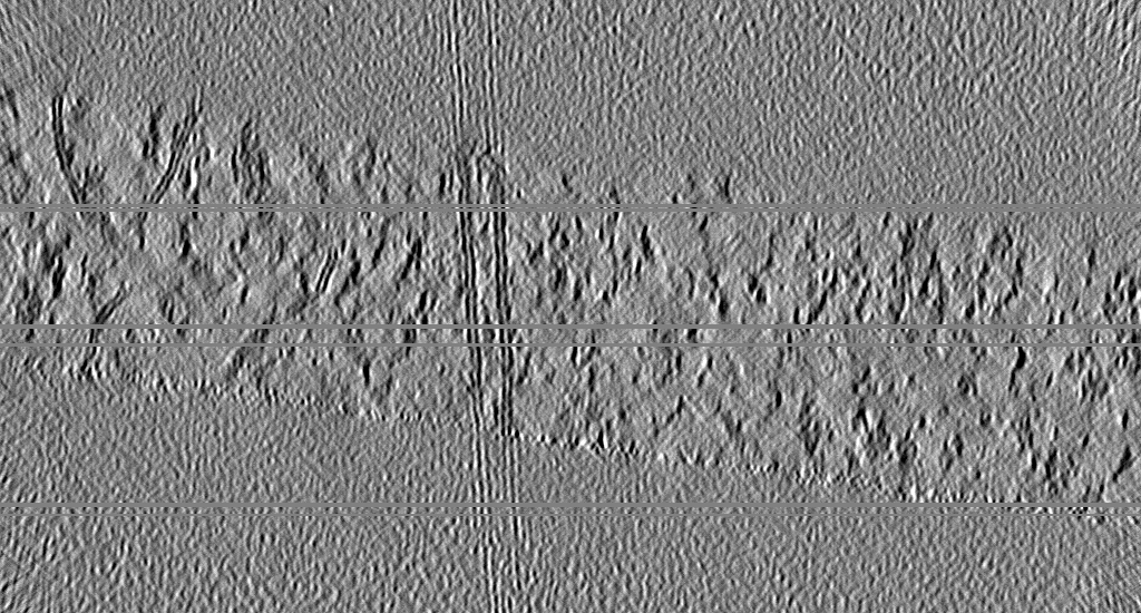
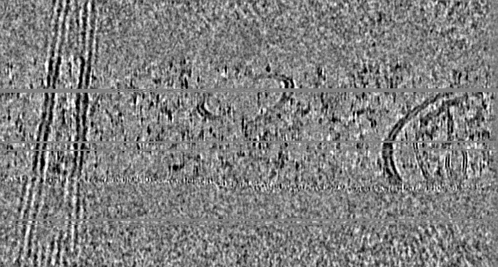
| タイトル | CryoCARE-denoised tomogram of two cholesterol-depleted HEK293 cells in close contact | |||||||||
 マップデータ マップデータ | CryoCARE-denoised tomogram (bin4) containing two HEK293 cells treated for 30 min with 10 mM MBCD in close contact. Visible features include both plasma membranes and the mitochondria of one cell. | |||||||||
 試料 試料 |
| |||||||||
 キーワード キーワード | plasma membrane / UNKNOWN FUNCTION | |||||||||
| 生物種 |  HEK293 (ヒト) HEK293 (ヒト) | |||||||||
| 手法 | 電子線トモグラフィー法 / クライオ電子顕微鏡法 | |||||||||
 データ登録者 データ登録者 | Glushkova D / Boehm S / Beck M | |||||||||
| 資金援助 |  ドイツ, 1件 ドイツ, 1件
| |||||||||
 引用 引用 | ジャーナル: J Cell Biol / 年: 2026 タイトル: Systematic membrane thickness variation across cellular organelles revealed by cryo-ET. 著者: Desislava Glushkova / Stefanie Böhm / Martin Beck / 要旨: In eukaryotes, membrane-bound organelles create distinct molecular environments. The compartmentalizing lipid bilayer is a dynamic composite material whose thickness and curvature modulate the ...In eukaryotes, membrane-bound organelles create distinct molecular environments. The compartmentalizing lipid bilayer is a dynamic composite material whose thickness and curvature modulate the structure and function of membrane proteins. In vitro, bilayer thickness correlates with lipid composition. Cellular membranes in situ, however, are continuously remodeled, and the spatial variation of their biophysical properties remains understudied. Here, we present a computational approach to measure local membrane thickness in cryo-electron tomograms. Our analysis of Chlamydomonas reinhardtii and human cells reveals systematic thickness variations within and across organelles. Notably, we observe thickness gradients across the Golgi apparatus that orthogonally support long-standing models of differential sorting of transmembrane proteins based on hydrophobic matching. Our publicly available workflow readily integrates within existing tomogram analysis pipelines and, when applied across experimental systems, provides a quantitative foundation for exploring relationships between membrane thickness and function in native cellular environments. | |||||||||
| 履歴 |
|
- 構造の表示
構造の表示
| 添付画像 |
|---|

- ダウンロードとリンク
-EMDBアーカイブ
| マップデータ | emd_55310.map.gz | 1.9 GB |  EMDBマップデータ形式 EMDBマップデータ形式 | |
|---|---|---|---|---|
| ヘッダ (付随情報) | emd-55310-v30.xmlemd-55310.xml | 10.5 KB 10.5 KB | 表示 表示 | EMDBヘッダ |
| 画像 |  emd_55310.png emd_55310.png | 263.1 KB | ||
| Filedesc metadata | emd-55310.cif.gz | 4 KB | ||
| アーカイブディレクトリ |  http://ftp.pdbj.org/pub/emdb/structures/EMD-55310ftp://ftp.pdbj.org/pub/emdb/structures/EMD-55310 http://ftp.pdbj.org/pub/emdb/structures/EMD-55310ftp://ftp.pdbj.org/pub/emdb/structures/EMD-55310 | HTTPS FTP |
-関連構造データ

-リンク
| EMDBのページ | EMDB (EBI/PDBe) / EMDataResource |
|---|
-マップ
| ファイル | ダウンロード / ファイル: emd_55310.map.gz / 形式: CCP4 / 大きさ: 2.1 GB / タイプ: IMAGE STORED AS FLOATING POINT NUMBER (4 BYTES) | ||||||||||||||||||||||||||||||||
|---|---|---|---|---|---|---|---|---|---|---|---|---|---|---|---|---|---|---|---|---|---|---|---|---|---|---|---|---|---|---|---|---|---|
| 注釈 | CryoCARE-denoised tomogram (bin4) containing two HEK293 cells treated for 30 min with 10 mM MBCD in close contact. Visible features include both plasma membranes and the mitochondria of one cell. | ||||||||||||||||||||||||||||||||
| 投影像・断面図 | 画像のコントロール
画像は Spider により作成 これらの図は立方格子座標系で作成されたものです | ||||||||||||||||||||||||||||||||
| ボクセルのサイズ | X=Y=Z: 7.884 Å | ||||||||||||||||||||||||||||||||
| 密度 |
| ||||||||||||||||||||||||||||||||
| 対称性 | 空間群: 1 | ||||||||||||||||||||||||||||||||
| 詳細 | EMDB XML:
|
 Z (Sec.)
 Y (Row.)
 X (Col.)


-添付データ
- 試料の構成要素
-全体 : CryoCARE-denoised tomogram of two cholesterol-depleted HEK293 cel...
| 全体 | 名称: CryoCARE-denoised tomogram of two cholesterol-depleted HEK293 cells in close contact |
|---|---|
| 要素 |
|
-超分子 #1: CryoCARE-denoised tomogram of two cholesterol-depleted HEK293 cel...
| 超分子 | 名称: CryoCARE-denoised tomogram of two cholesterol-depleted HEK293 cells in close contact タイプ: cell / ID: 1 / 親要素: 0 |
|---|---|
| 由来(天然) | 生物種: HEK293 (ヒト) |
-実験情報
-構造解析
| 手法 | クライオ電子顕微鏡法 |
|---|---|
 解析 解析 | 電子線トモグラフィー法 |
| 試料の集合状態 | cell |
-試料調製
| 緩衝液 | pH: 7.4 / 構成要素 - 濃度: 10.0 mM / 構成要素 - 名称: Methyl-beta-cyclodextrin |
|---|---|
| グリッド | モデル: Quantifoil R2/2 / 材質: GOLD / メッシュ: 200 / 支持フィルム - 材質: SILICON DIOXIDE / 支持フィルム - トポロジー: HOLEY / 前処理 - タイプ: GLOW DISCHARGE / 前処理 - 時間: 90 sec. |
| 凍結 | 凍結剤: ETHANE / 装置: LEICA EM GP |
| 切片作成 | 集束イオンビーム - 装置: OTHER / 集束イオンビーム - イオン: OTHER / 集束イオンビーム - 電圧: 30 / 集束イオンビーム - 電流: 0.1 / 集束イオンビーム - 時間: 3600 / 集束イオンビーム - 温度: 80 K / 集束イオンビーム - Initial thickness: 1000 / 集束イオンビーム - 最終 厚さ: 200 集束イオンビーム - 詳細: The value given for _em_focused_ion_beam.instrument is Aquilos 2 FIB-SEM. This is not in a list of allowed values {'DB235', 'OTHER'} so OTHER is written into the XML file. |
- 電子顕微鏡法
| 顕微鏡 | TFS KRIOS |
|---|---|
| 撮影 | フィルム・検出器のモデル: FEI FALCON IV (4k x 4k) 平均電子線量: 2.0 e/Å2 |
| 電子線 | 加速電圧: 300 kV / 電子線源:  FIELD EMISSION GUN FIELD EMISSION GUN |
| 電子光学系 | 照射モード: FLOOD BEAM / 撮影モード: BRIGHT FIELD / 最大 デフォーカス(公称値): 3.5 µm / 最小 デフォーカス(公称値): 1.5 µm |
| 実験機器 |  モデル: Titan Krios / 画像提供: FEI Company |
-画像解析
| 最終 再構成 | ソフトウェア - 名称: IMOD / 使用した粒子像数: 61 |
|---|---|
| CTF補正 | タイプ: NONE |
