 Movie
Movie Controller

[English] 日本語
 Yorodumi
- EMDB-55310: CryoCARE-denoised tomogram of two cholesterol-depleted HEK293 cel... -
+ Open data

- Basic information
| Entry |  | |||||||||
|---|---|---|---|---|---|---|---|---|---|---|
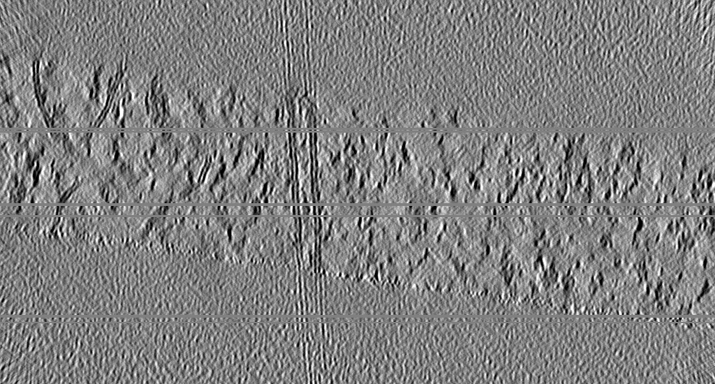
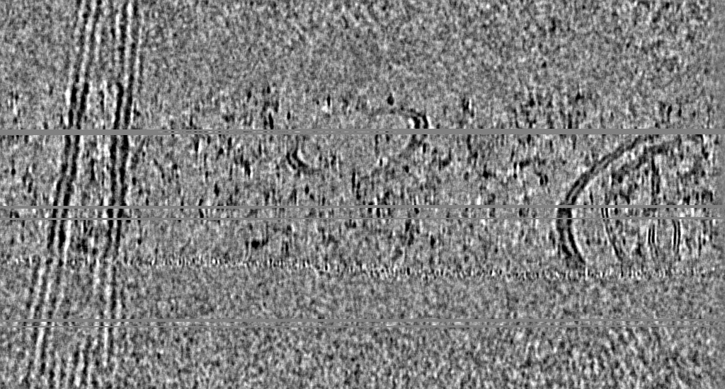
| Title | CryoCARE-denoised tomogram of two cholesterol-depleted HEK293 cells in close contact | |||||||||
 Map data Map data | CryoCARE-denoised tomogram (bin4) containing two HEK293 cells treated for 30 min with 10 mM MBCD in close contact. Visible features include both plasma membranes and the mitochondria of one cell. | |||||||||
 Sample Sample |
| |||||||||
 Keywords Keywords | plasma membrane / UNKNOWN FUNCTION | |||||||||
| Biological species |  HEK293 (human) HEK293 (human) | |||||||||
| Method | electron tomography / cryo EM | |||||||||
 Authors Authors | Glushkova D / Boehm S / Beck M | |||||||||
| Funding support |  Germany, 1 items Germany, 1 items
| |||||||||
 Citation Citation | Journal: J Cell Biol / Year: 2026 Title: Systematic membrane thickness variation across cellular organelles revealed by cryo-ET. Authors: Desislava Glushkova / Stefanie Böhm / Martin Beck / Abstract: In eukaryotes, membrane-bound organelles create distinct molecular environments. The compartmentalizing lipid bilayer is a dynamic composite material whose thickness and curvature modulate the ...In eukaryotes, membrane-bound organelles create distinct molecular environments. The compartmentalizing lipid bilayer is a dynamic composite material whose thickness and curvature modulate the structure and function of membrane proteins. In vitro, bilayer thickness correlates with lipid composition. Cellular membranes in situ, however, are continuously remodeled, and the spatial variation of their biophysical properties remains understudied. Here, we present a computational approach to measure local membrane thickness in cryo-electron tomograms. Our analysis of Chlamydomonas reinhardtii and human cells reveals systematic thickness variations within and across organelles. Notably, we observe thickness gradients across the Golgi apparatus that orthogonally support long-standing models of differential sorting of transmembrane proteins based on hydrophobic matching. Our publicly available workflow readily integrates within existing tomogram analysis pipelines and, when applied across experimental systems, provides a quantitative foundation for exploring relationships between membrane thickness and function in native cellular environments. | |||||||||
| History |
|
- Structure visualization
| Supplemental images |
|---|

- Downloads & links
-EMDB archive
| Map data | emd_55310.map.gz | 1.9 GB |  EMDB map data format EMDB map data format | |
|---|---|---|---|---|
| Header (meta data) | emd-55310-v30.xmlemd-55310.xml | 10.5 KB 10.5 KB | Display Display | EMDB header |
| Images |  emd_55310.png emd_55310.png | 263.1 KB | ||
| Filedesc metadata | emd-55310.cif.gz | 4 KB | ||
| Archive directory |  http://ftp.pdbj.org/pub/emdb/structures/EMD-55310ftp://ftp.pdbj.org/pub/emdb/structures/EMD-55310 http://ftp.pdbj.org/pub/emdb/structures/EMD-55310ftp://ftp.pdbj.org/pub/emdb/structures/EMD-55310 | HTTPS FTP |
-Related structure data

-Links
| EMDB pages | EMDB (EBI/PDBe) / EMDataResource |
|---|
-Map
| File | Download / File: emd_55310.map.gz / Format: CCP4 / Size: 2.1 GB / Type: IMAGE STORED AS FLOATING POINT NUMBER (4 BYTES) | ||||||||||||||||||||||||||||||||
|---|---|---|---|---|---|---|---|---|---|---|---|---|---|---|---|---|---|---|---|---|---|---|---|---|---|---|---|---|---|---|---|---|---|
| Annotation | CryoCARE-denoised tomogram (bin4) containing two HEK293 cells treated for 30 min with 10 mM MBCD in close contact. Visible features include both plasma membranes and the mitochondria of one cell. | ||||||||||||||||||||||||||||||||
| Projections & slices | Image control
Images are generated by Spider. generated in cubic-lattice coordinate | ||||||||||||||||||||||||||||||||
| Voxel size | X=Y=Z: 7.884 Å | ||||||||||||||||||||||||||||||||
| Density |
| ||||||||||||||||||||||||||||||||
| Symmetry | Space group: 1 | ||||||||||||||||||||||||||||||||
| Details | EMDB XML:
|
 Z (Sec.)
 Y (Row.)
 X (Col.)


-Supplemental data
- Sample components
-Entire : CryoCARE-denoised tomogram of two cholesterol-depleted HEK293 cel...
| Entire | Name: CryoCARE-denoised tomogram of two cholesterol-depleted HEK293 cells in close contact |
|---|---|
| Components |
|
-Supramolecule #1: CryoCARE-denoised tomogram of two cholesterol-depleted HEK293 cel...
| Supramolecule | Name: CryoCARE-denoised tomogram of two cholesterol-depleted HEK293 cells in close contact type: cell / ID: 1 / Parent: 0 |
|---|---|
| Source (natural) | Organism: HEK293 (human) |
-Experimental details
-Structure determination
| Method | cryo EM |
|---|---|
 Processing Processing | electron tomography |
| Aggregation state | cell |
-Sample preparation
| Buffer | pH: 7.4 / Component - Concentration: 10.0 mM / Component - Name: Methyl-beta-cyclodextrin |
|---|---|
| Grid | Model: Quantifoil R2/2 / Material: GOLD / Mesh: 200 / Support film - Material: SILICON DIOXIDE / Support film - topology: HOLEY / Pretreatment - Type: GLOW DISCHARGE / Pretreatment - Time: 90 sec. |
| Vitrification | Cryogen name: ETHANE / Instrument: LEICA EM GP |
| Sectioning | Focused ion beam - Instrument: OTHER / Focused ion beam - Ion: OTHER / Focused ion beam - Voltage: 30 / Focused ion beam - Current: 0.1 / Focused ion beam - Duration: 3600 / Focused ion beam - Temperature: 80 K / Focused ion beam - Initial thickness: 1000 / Focused ion beam - Final thickness: 200 Focused ion beam - Details: The value given for _em_focused_ion_beam.instrument is Aquilos 2 FIB-SEM. This is not in a list of allowed values {'DB235', 'OTHER'} so OTHER is written into the XML file. |
- Electron microscopy
| Microscope | TFS KRIOS |
|---|---|
| Image recording | Film or detector model: FEI FALCON IV (4k x 4k) / Average electron dose: 2.0 e/Å2 |
| Electron beam | Acceleration voltage: 300 kV / Electron source:  FIELD EMISSION GUN FIELD EMISSION GUN |
| Electron optics | Illumination mode: FLOOD BEAM / Imaging mode: BRIGHT FIELD / Nominal defocus max: 3.5 µm / Nominal defocus min: 1.5 µm |
| Experimental equipment |  Model: Titan Krios / Image courtesy: FEI Company |
-Image processing
| Final reconstruction | Software - Name: IMOD / Number images used: 61 |
|---|---|
| CTF correction | Type: NONE |
