 ムービー
ムービー コントローラー


+ データを開く

- 基本情報
基本情報
| 登録情報 | データベース: EMDB / ID: EMD-5528 | |||||||||
|---|---|---|---|---|---|---|---|---|---|---|
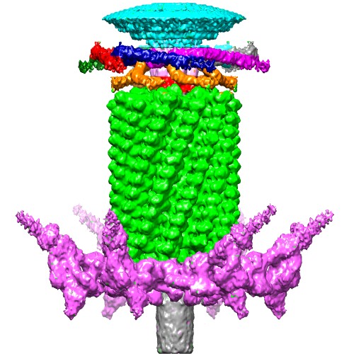
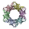
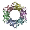
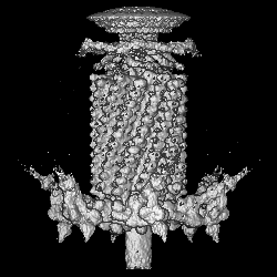
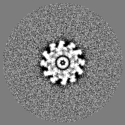
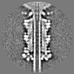
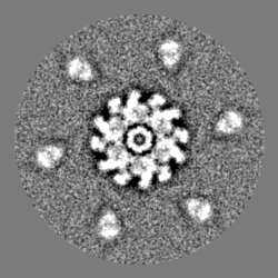
| タイトル | Cryo-EM structure of the contracted bacteriophage T4 tail containing the collar and whiskers made of fibritin molecules. | |||||||||
 マップデータ マップデータ | Cryo-EM reconstruction of the contracted T4 tail containing the collar and whiskers | |||||||||
 試料 試料 |
| |||||||||
 キーワード キーワード | bacteriophage / T4 / phage / collar / whiskers / fibritin | |||||||||
| 機能・相同性 | Fibritin C-terminal / Fibritin C-terminal region / virion component / Fibritin 機能・相同性情報 機能・相同性情報 | |||||||||
| 生物種 |  Enterobacteria phage T4 (ファージ) Enterobacteria phage T4 (ファージ) | |||||||||
| 手法 | 単粒子再構成法 / クライオ電子顕微鏡法 / 解像度: 25.0 Å | |||||||||
 データ登録者 データ登録者 | Fokine A / Zhang Z / Kanamaru S / Bowman VD / Aksyuk AA / Arisaka F / Rao VB / Rossmann MG | |||||||||
 引用 引用 | ジャーナル: J Mol Biol / 年: 2013 タイトル: The molecular architecture of the bacteriophage T4 neck. 著者: Andrei Fokine / Zhihong Zhang / Shuji Kanamaru / Valorie D Bowman / Anastasia A Aksyuk / Fumio Arisaka / Venigalla B Rao / Michael G Rossmann /  要旨: A hexamer of the bacteriophage T4 tail terminator protein, gp15, attaches to the top of the phage tail stabilizing the contractile sheath and forming the interface for binding of the independently ...A hexamer of the bacteriophage T4 tail terminator protein, gp15, attaches to the top of the phage tail stabilizing the contractile sheath and forming the interface for binding of the independently assembled head. Here we report the crystal structure of the gp15 hexamer, describe its interactions in T4 virions that have either an extended tail or a contracted tail, and discuss its structural relationship to other phage proteins. The neck of T4 virions is decorated by the "collar" and "whiskers", made of fibritin molecules. Fibritin acts as a chaperone helping to attach the long tail fibers to the virus during the assembly process. The collar and whiskers are environment-sensing devices, regulating the retraction of the long tail fibers under unfavorable conditions, thus preventing infection. Cryo-electron microscopy analysis suggests that twelve fibritin molecules attach to the phage neck with six molecules forming the collar and six molecules forming the whiskers. | |||||||||
| 履歴 |
|
- 構造の表示
構造の表示
| ムービー |
ムービービューア |
|---|---|
| 構造ビューア | EMマップ: SurfViewMolmilJmol/JSmol |
| 添付画像 |
- ダウンロードとリンク
-EMDBアーカイブ
| マップデータ | emd_5528.map.gz | 26 MB | EMDBマップデータ形式 | |
|---|---|---|---|---|
| ヘッダ (付随情報) | emd-5528-v30.xmlemd-5528.xml | 9.3 KB 9.3 KB | 表示 表示 | EMDBヘッダ |
| 画像 |  emd_5528_1.jpg emd_5528_1.jpg | 73.5 KB | ||
| アーカイブディレクトリ |  http://ftp.pdbj.org/pub/emdb/structures/EMD-5528ftp://ftp.pdbj.org/pub/emdb/structures/EMD-5528 http://ftp.pdbj.org/pub/emdb/structures/EMD-5528ftp://ftp.pdbj.org/pub/emdb/structures/EMD-5528 | HTTPS FTP |
-検証レポート
| 文書・要旨 | emd_5528_validation.pdf.gz | 376.8 KB | 表示 | EMDB検証レポート |
|---|---|---|---|---|
| 文書・詳細版 | emd_5528_full_validation.pdf.gz | 376.3 KB | 表示 | |
| XML形式データ | emd_5528_validation.xml.gz | 6.4 KB | 表示 | |
| アーカイブディレクトリ | https://ftp.pdbj.org/pub/emdb/validation_reports/EMD-5528ftp://ftp.pdbj.org/pub/emdb/validation_reports/EMD-5528 | HTTPS FTP |
-関連構造データ
-リンク
| EMDBのページ | EMDB (EBI/PDBe) / EMDataResource |
|---|---|
| 「今月の分子」の関連する項目 |


-マップ
| ファイル | ダウンロード / ファイル: emd_5528.map.gz / 形式: CCP4 / 大きさ: 58.2 MB / タイプ: IMAGE STORED AS FLOATING POINT NUMBER (4 BYTES) | ||||||||||||||||||||||||||||||||||||||||||||||||||||||||||||||||||||
|---|---|---|---|---|---|---|---|---|---|---|---|---|---|---|---|---|---|---|---|---|---|---|---|---|---|---|---|---|---|---|---|---|---|---|---|---|---|---|---|---|---|---|---|---|---|---|---|---|---|---|---|---|---|---|---|---|---|---|---|---|---|---|---|---|---|---|---|---|---|
| 注釈 | Cryo-EM reconstruction of the contracted T4 tail containing the collar and whiskers | ||||||||||||||||||||||||||||||||||||||||||||||||||||||||||||||||||||
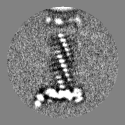
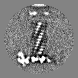
| 投影像・断面図 | 画像のコントロール
画像は Spider により作成 | ||||||||||||||||||||||||||||||||||||||||||||||||||||||||||||||||||||
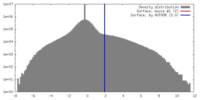
| ボクセルのサイズ | X=Y=Z: 3.572 Å | ||||||||||||||||||||||||||||||||||||||||||||||||||||||||||||||||||||
| 密度 |
| ||||||||||||||||||||||||||||||||||||||||||||||||||||||||||||||||||||
| 対称性 | 空間群: 1 | ||||||||||||||||||||||||||||||||||||||||||||||||||||||||||||||||||||
| 詳細 | EMDB XML:
CCP4マップ ヘッダ情報:
| ||||||||||||||||||||||||||||||||||||||||||||||||||||||||||||||||||||
 Z (Sec.)
 Y (Row.)
 X (Col.)


-添付データ
- 試料の構成要素
-全体 : bacteriophage T4
| 全体 | 名称: bacteriophage T4 (ファージ) |
|---|---|
| 要素 |
|
-超分子 #1000: bacteriophage T4
| 超分子 | 名称: bacteriophage T4 / タイプ: sample / ID: 1000 / 詳細: The phage tails were contracted using 4M urea. / Number unique components: 1 |
|---|
-超分子 #1: Enterobacteria phage T4
| 超分子 | 名称: Enterobacteria phage T4 / タイプ: virus / ID: 1 / Name.synonym: Enterobacteria phage T4 / NCBI-ID: 10665 / 生物種: Enterobacteria phage T4 / データベース: NCBI / ウイルスタイプ: VIRION / ウイルス・単離状態: STRAIN / ウイルス・エンベロープ: No / ウイルス・中空状態: No / Syn species name: Enterobacteria phage T4 |
|---|---|
| 宿主 | 生物種:  |
-実験情報
-構造解析
| 手法 | クライオ電子顕微鏡法 |
|---|---|
 解析 解析 | 単粒子再構成法 |
| 試料の集合状態 | particle |
-試料調製
| 緩衝液 | pH: 8 / 詳細: 50 mM Tris-HCl, pH 8.0, 0.2 M NaCl, 8 mM MgCl2 |
|---|---|
| 凍結 | 凍結剤: ETHANE / 装置: HOMEMADE PLUNGER |
- 電子顕微鏡法
| 顕微鏡 | FEI/PHILIPS CM200FEG |
|---|---|
| 日付 | 2007年3月7日 |
| 撮影 | カテゴリ: FILM / フィルム・検出器のモデル: KODAK SO-163 FILM / デジタル化 - スキャナー: ZEISS SCAI / デジタル化 - サンプリング間隔: 7 µm / 実像数: 97 / 平均電子線量: 16 e/Å2 / ビット/ピクセル: 8 |
| Tilt angle min | 0 |
| Tilt angle max | 0 |
| 電子線 | 加速電圧: 200 kV / 電子線源:  FIELD EMISSION GUN FIELD EMISSION GUN |
| 電子光学系 | 倍率(補正後): 39190 / 照射モード: SPOT SCAN / 撮影モード: BRIGHT FIELD / Cs: 2 mm / 最大 デフォーカス(公称値): 3.0 µm / 最小 デフォーカス(公称値): 1.5 µm / 倍率(公称値): 38000 |
| 試料ステージ | 試料ホルダーモデル: GATAN LIQUID NITROGEN |
-画像解析
| CTF補正 | 詳細: phase flipping |
|---|---|
| 最終 再構成 | アルゴリズム: OTHER / 解像度のタイプ: BY AUTHOR / 解像度: 25.0 Å / 解像度の算出法: FSC 0.5 CUT-OFF / ソフトウェア - 名称: EMAN 詳細: The reconstruction of the contracted T4 tail which lacked the collar and whiskers (EMDB accession code 1086; Leiman et al., 2004, Cell 118, 419-429) was used as the initial model. 使用した粒子像数: 2727 |
