 Movie
Movie Controller

[English] 日本語
 Yorodumi
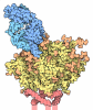
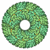
- EMDB-54918: Prefusion-stabilized Nipah virus fusion protein in complex with i... -
+ Open data

- Basic information
| Entry |  | ||||||||||||
|---|---|---|---|---|---|---|---|---|---|---|---|---|---|
| Title | Prefusion-stabilized Nipah virus fusion protein in complex with inhibitory nanobody F112 | ||||||||||||
 Map data Map data | |||||||||||||
 Sample Sample |
| ||||||||||||
 Keywords Keywords | Fusion protein / antiviral / nanobody / VIRAL PROTEIN | ||||||||||||
| Function / homology | membrane fusion involved in viral entry into host cell / Precursor fusion glycoprotein F0, Paramyxoviridae / Fusion glycoprotein F0 / fusion of virus membrane with host plasma membrane / viral envelope / symbiont entry into host cell / host cell plasma membrane / virion membrane / Fusion glycoprotein F0 Function and homology information Function and homology information | ||||||||||||
| Biological species |  Henipavirus nipahense / Henipavirus nipahense /   Vicugna pacos (alpaca) Vicugna pacos (alpaca) | ||||||||||||
| Method | single particle reconstruction / cryo EM / Resolution: 2.79 Å | ||||||||||||
 Authors Authors | Kralova A / Hanke L | ||||||||||||
| Funding support | European Union,  Sweden, 3 items Sweden, 3 items
| ||||||||||||
 Citation Citation | Journal: To Be Published Title: Prefusion-stabilized Nipah virus fusion protein in complex with inhibitory nanobody F112 Authors: Kralova A / Hanke L | ||||||||||||
| History |
|
- Structure visualization
| Supplemental images |
|---|

- Downloads & links
-EMDB archive
| Map data | emd_54918.map.gz | 89.7 MB | EMDB map data format | |
|---|---|---|---|---|
| Header (meta data) | emd-54918-v30.xmlemd-54918.xml | 18.3 KB 18.3 KB | Display Display | EMDB header |
| FSC (resolution estimation) | emd_54918_fsc.xml | 16.4 KB | Display | FSC data file |
| Images |  emd_54918.png emd_54918.png | 67.6 KB | ||
| Filedesc metadata | emd-54918.cif.gz | 6.1 KB | ||
| Others | emd_54918_half_map_1.map.gzemd_54918_half_map_2.map.gz | 165.4 MB 165.4 MB | ||
| Archive directory |  https://data.pdbj.org/pub/emdb/structures/EMD-54918ftp://data.pdbj.org/pub/emdb/structures/EMD-54918 https://data.pdbj.org/pub/emdb/structures/EMD-54918ftp://data.pdbj.org/pub/emdb/structures/EMD-54918 | HTTPS FTP |
-Related structure data
| Related structure data |  9shwMC M: atomic model generated by this map C: citing same article ( |
|---|---|
| Similar structure data |
-Links
| EMDB pages | EMDB (EBI/PDBe) / EMDataResource |
|---|---|
| Related items in Molecule of the Month |
-Map
| File | Download / File: emd_54918.map.gz / Format: CCP4 / Size: 178 MB / Type: IMAGE STORED AS FLOATING POINT NUMBER (4 BYTES) | ||||||||||||||||||||||||||||||||||||
|---|---|---|---|---|---|---|---|---|---|---|---|---|---|---|---|---|---|---|---|---|---|---|---|---|---|---|---|---|---|---|---|---|---|---|---|---|---|
| Projections & slices | Image control
Images are generated by Spider. | ||||||||||||||||||||||||||||||||||||
| Voxel size | X=Y=Z: 0.828 Å | ||||||||||||||||||||||||||||||||||||
| Density |
| ||||||||||||||||||||||||||||||||||||
| Symmetry | Space group: 1 | ||||||||||||||||||||||||||||||||||||
| Details | EMDB XML:
|
 Z (Sec.)
 Y (Row.)
 X (Col.)


-Supplemental data
-Half map: #2
| File | emd_54918_half_map_1.map | ||||||||||||
|---|---|---|---|---|---|---|---|---|---|---|---|---|---|
| Projections & Slices |
| ||||||||||||
| Density Histograms |


-Half map: #1
| File | emd_54918_half_map_2.map | ||||||||||||
|---|---|---|---|---|---|---|---|---|---|---|---|---|---|
| Projections & Slices |
| ||||||||||||
| Density Histograms |


- Sample components
-Entire : Prefusion-stabilized Nipah virus F glycoprotein in complex with t...
| Entire | Name: Prefusion-stabilized Nipah virus F glycoprotein in complex with the neutralizing F112 nanobody |
|---|---|
| Components |
|
-Supramolecule #1: Prefusion-stabilized Nipah virus F glycoprotein in complex with t...
| Supramolecule | Name: Prefusion-stabilized Nipah virus F glycoprotein in complex with the neutralizing F112 nanobody type: complex / ID: 1 / Parent: 0 / Macromolecule list: all |
|---|---|
| Source (natural) | Organism: Henipavirus nipahense |
| Molecular weight | Theoretical: 44 KDa |
-Supramolecule #2: Fusion glycoprotein F
| Supramolecule | Name: Fusion glycoprotein F / type: complex / ID: 2 / Parent: 1 / Macromolecule list: #1 |
|---|---|
| Source (natural) | Organism: Henipavirus nipahense |
-Supramolecule #3: F112 nanobody
| Supramolecule | Name: F112 nanobody / type: complex / ID: 3 / Parent: 2 / Macromolecule list: #2 |
|---|---|
| Source (natural) | Organism: Vicugna pacos (alpaca) |
-Macromolecule #1: Fusion glycoprotein F0
| Macromolecule | Name: Fusion glycoprotein F0 / type: protein_or_peptide / ID: 1 / Number of copies: 3 / Enantiomer: LEVO |
|---|---|
| Source (natural) | Organism: Henipavirus nipahense |
| Molecular weight | Theoretical: 62.510625 KDa |
| Recombinant expression | Organism:  Homo sapiens (human) Homo sapiens (human) |
| Sequence | String: MVVILDKRCY CNLLILILMI SECSVGILHY EKLSKIGLVK GVTRKYKIKS NPLTKDIVIK MIPNVSNMSQ CTGSVMENYK TRLNGILTP IKGALEIYKN NTHDCVGDVR LAGVCMAGVA IGIATAAQIT AGVALYEAMK NADNINKLKS SIESTNEAVV K LQETAEKT ...String: MVVILDKRCY CNLLILILMI SECSVGILHY EKLSKIGLVK GVTRKYKIKS NPLTKDIVIK MIPNVSNMSQ CTGSVMENYK TRLNGILTP IKGALEIYKN NTHDCVGDVR LAGVCMAGVA IGIATAAQIT AGVALYEAMK NADNINKLKS SIESTNEAVV K LQETAEKT VYVFTALQDY INTNLVPTID KIPCKQTELS LDLALSKYLS DLLFVFGPNL QDPVSNSMTI QAISQAFGGN YE TLLRTLG YATEDFDDLL ESDSITGQII YVDLSSYYII VRVYFPILTE IQQAYIQELL PVSFNNDDSE WISIVPNFIL VRN TLISNI EIGFCLITKR SVICNQDYAT PMTNNMRECL TGSTEKCPRE LVVSSHVPRF ALSNGVLFAN CISVTCQCQT TGRA ISQSG EQTLLMIDNT TCPTAVLGNV IISLGKYLGS VNYNSEGIAI GPPVFTDKVD ISSQISSMNQ SLQQSKDYIK EAQRL LDTV NPSMKQIEDK IEEILSKIYH IENEIARIKK LIGEAPGGIE GRKLHHHHHH HHSAWSHPQF EKGGGSGGGG SGGSAW SHP QFEK UniProtKB: Fusion glycoprotein F0 |
-Macromolecule #2: F112 nanobody
| Macromolecule | Name: F112 nanobody / type: protein_or_peptide / ID: 2 / Number of copies: 3 / Enantiomer: LEVO |
|---|---|
| Source (natural) | Organism: Vicugna pacos (alpaca) |
| Molecular weight | Theoretical: 14.323876 KDa |
| Recombinant expression | Organism:   Escherichia coli BL21 (bacteria) Escherichia coli BL21 (bacteria) |
| Sequence | String: QVQLVESGGG LVQPGGSLRL SCAVLGSIYG INAMAWYRRA PGSQRELVAV GSGDRINYAD AVKGRFTISR DDAKNSVYLQ MNSLKPEDT AVYYCQANIN TSGGWFREYW GQGTQVTVSS GGLPGTGGHH HHHH |
-Experimental details
-Structure determination
| Method | cryo EM |
|---|---|
 Processing Processing | single particle reconstruction |
| Aggregation state | 3D array |
-Sample preparation
| Concentration | 0.24 mg/mL | ||||||||||||
|---|---|---|---|---|---|---|---|---|---|---|---|---|---|
| Buffer | pH: 8 Component:
| ||||||||||||
| Vitrification | Cryogen name: ETHANE / Chamber humidity: 100 % / Chamber temperature: 295.15 K / Instrument: FEI VITROBOT MARK IV |
- Electron microscopy
| Microscope | TFS KRIOS |
|---|---|
| Image recording | Film or detector model: GATAN K3 (6k x 4k) / Average electron dose: 48.28 e/Å2 |
| Electron beam | Acceleration voltage: 300 kV / Electron source:  FIELD EMISSION GUN FIELD EMISSION GUN |
| Electron optics | Illumination mode: FLOOD BEAM / Imaging mode: BRIGHT FIELD / Nominal defocus max: 1.7 µm / Nominal defocus min: 0.5 µm |
| Experimental equipment |  Model: Titan Krios / Image courtesy: FEI Company |
+Image processing

-Atomic model buiding 1
| Initial model | Chain - Source name: AlphaFold |
|---|---|
| Output model | PDB-9shw: |
