- EMDB-54787: Cryo-EM structure of PfHT1 bound to 2,5-anhydro-D-mannitol -
+
Open data
ID or keywords:
Loading...
-
Basic information
Entry
Database: EMDB / ID: EMD-54787
Title
Cryo-EM structure of PfHT1 bound to 2,5-anhydro-D-mannitol
Map data
Sample
Complex: PfHT1 dimer with 2,5-anhydro-D-mannitol
Protein or peptide: Hexose transporter 1,Green fluorescent protein
Ligand: 2.5-anhydro-D-mannitol
Keywords
Sugar transporter / Plasmodium falciparum hexose transporter 1 / TRANSPORT PROTEIN
Function / homology
Function and homology information
hexose transmembrane transporter activity / bioluminescence / generation of precursor metabolites and energy / plasma membrane Similarity search - Function
Glucose transporter GLUT / Sugar/inositol transporter / Sugar transport proteins signature 2. / Sugar transport proteins signature 1. / Sugar transporter, conserved site / Major facilitator, sugar transporter-like / Sugar (and other) transporter / Major facilitator superfamily domain / Major facilitator superfamily (MFS) profile. / MFS transporter superfamily ...Glucose transporter GLUT / Sugar/inositol transporter / Sugar transport proteins signature 2. / Sugar transport proteins signature 1. / Sugar transporter, conserved site / Major facilitator, sugar transporter-like / Sugar (and other) transporter / Major facilitator superfamily domain / Major facilitator superfamily (MFS) profile. / MFS transporter superfamily / Green fluorescent protein, GFP / Green fluorescent protein-related / Green fluorescent protein / Green fluorescent protein Similarity search - Domain/homology
Journal: Nat Struct Mol Biol / Year: 2026 Title: A two-step mechanism for sugar translocation. Authors: Do-Hwan Ahn / Claudia Alleva / Tom Reichenbach / Ashutosh Gulati / Alessandro Ruda / Marta Bonaccorsi / Jakob M Silberberg / Magnus Claesson / Albert Suades / Lucie Delemotte / Göran Widmalm / David Drew / Abstract: In mammals, glucose transporters (GLUTs) mediate organism-wide sugar distribution, yet the molecular basis of substrate specificity remains unclear. The bacterial xylose transporter XylE serves as a ...In mammals, glucose transporters (GLUTs) mediate organism-wide sugar distribution, yet the molecular basis of substrate specificity remains unclear. The bacterial xylose transporter XylE serves as a model for GLUTs. However, although xylose and glucose bind with a similar affinity, xylose is transported, but glucose acts as an inhibitor. Here, using saturation transfer difference (STD) nuclear magnetic resonance (NMR) spectroscopy, we distinguished transported sugars from sugar inhibitors. Our findings revealed that only transported sugars generate STD NMR signals, which are abolished for xylose when XylE is trapped in either outward- or inward-facing conformations. Engineering the sugar-binding pocket and gating helix TM7b enabled glucose transport by XylE and corresponding STD signals. Using complementary molecular dynamics simulations, together with structural, biochemical and STD NMR analysis of related parasitic and mammalian GLUTs, we identified TM7b as a key determinant of occluded state formation. We conclude that, rather than the initial substrate-binding event observed in experimental structures, formation of a substrate-induced transition-state intermediate is the primary determinant of specificity in transporters.
In the structure databanks used in Yorodumi, some data are registered as the other names, "COVID-19 virus" and "2019-nCoV". Here are the details of the virus and the list of structure data.
Jan 31, 2019. EMDB accession codes are about to change! (news from PDBe EMDB page)
EMDB accession codes are about to change! (news from PDBe EMDB page)
The allocation of 4 digits for EMDB accession codes will soon come to an end. Whilst these codes will remain in use, new EMDB accession codes will include an additional digit and will expand incrementally as the available range of codes is exhausted. The current 4-digit format prefixed with “EMD-” (i.e. EMD-XXXX) will advance to a 5-digit format (i.e. EMD-XXXXX), and so on. It is currently estimated that the 4-digit codes will be depleted around Spring 2019, at which point the 5-digit format will come into force.
The EM Navigator/Yorodumi systems omit the EMD- prefix.
Related info.:Q: What is EMD? / ID/Accession-code notation in Yorodumi/EM Navigator
Yorodumi is a browser for structure data from EMDB, PDB, SASBDB, etc.
This page is also the successor to EM Navigator detail page, and also detail information page/front-end page for Omokage search.
The word "yorodu" (or yorozu) is an old Japanese word meaning "ten thousand". "mi" (miru) is to see.
Related info.:EMDB / PDB / SASBDB / Comparison of 3 databanks / Yorodumi Search / Aug 31, 2016. New EM Navigator & Yorodumi / Yorodumi Papers / Jmol/JSmol / Function and homology information / Changes in new EM Navigator and Yorodumi
 Movie
Movie Controller


 Open data
 Basic information
 Map data
 Sample
 Keywords
 Function and homology information

 Aequorea victoria (jellyfish)
 Authors
 Sweden,
  Denmark, 3 items
  Citation
 Structure visualization
 Downloads & links
 emd_54787.png
 http://ftp.pdbj.org/pub/emdb/structures/EMD-54787
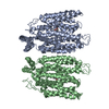


 Z (Sec.)
 Y (Row.)
 X (Col.)


 Sample components
 Processing
 Electron microscopy
 FIELD EMISSION GUN

