 Movie
Movie Controller


+ Open data

- Basic information
| Entry | Database: EMDB / ID: EMD-5444 | |||||||||
|---|---|---|---|---|---|---|---|---|---|---|
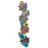
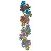
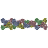
| Title | MDA5-dsRNA filament | |||||||||
 Map data Map data | reconstruction of MDA5-dsRNA | |||||||||
 Sample Sample |
| |||||||||
 Keywords Keywords | MAVS nucleation / nucleoprotein filament / helical polymer | |||||||||
| Biological species |  | |||||||||
| Method | helical reconstruction / Resolution: 20.0 Å | |||||||||
 Authors Authors | Berke IC / Yu X / Modis Y / Egelman EH | |||||||||
 Citation Citation | Journal: Proc Natl Acad Sci U S A / Year: 2012 Title: MDA5 assembles into a polar helical filament on dsRNA. Authors: Ian C Berke / Xiong Yu / Yorgo Modis / Edward H Egelman /  Abstract: Melanoma differentiation-associated protein 5 (MDA5) detects viral dsRNA in the cytoplasm. On binding of RNA, MDA5 forms polymers, which trigger assembly of the signaling adaptor mitochondrial ...Melanoma differentiation-associated protein 5 (MDA5) detects viral dsRNA in the cytoplasm. On binding of RNA, MDA5 forms polymers, which trigger assembly of the signaling adaptor mitochondrial antiviral-signaling protein (MAVS) into its active fibril form. The molecular mechanism of MDA5 signaling is not well understood, however. Here we show that MDA5 forms helical filaments on dsRNA and report the 3D structure of the filaments using electron microscopy (EM) and image reconstruction. MDA5 assembles into a polar, single-start helix around the RNA. Fitting of an MDA5 homology model into the structure suggests a key role for the MDA5 C-terminal domain in cooperative filament assembly. Our study supports a signal transduction mechanism in which the helical array of MDA5 within filaments nucleates the assembly of MAVS fibrils. We conclude that MDA5 is a polymerization-dependent signaling platform that uses the amyloid-like self-propagating properties of MAVS to amplify signaling. | |||||||||
| History |
|
- Structure visualization
| Movie |
 Movie viewer Movie viewer |
|---|---|
| Structure viewer | EM map: SurfViewMolmilJmol/JSmol |
| Supplemental images |
 UCSF Chimera
- Downloads & links
-EMDB archive
| Map data | emd_5444.map.gz | 314.1 KB | EMDB map data format | |
|---|---|---|---|---|
| Header (meta data) | emd-5444-v30.xmlemd-5444.xml | 7.4 KB 7.4 KB | Display Display | EMDB header |
| Images | 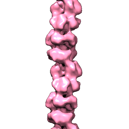 emd_5444_1.png emd_5444_1.png | 89.9 KB | ||
| Archive directory |  http://ftp.pdbj.org/pub/emdb/structures/EMD-5444ftp://ftp.pdbj.org/pub/emdb/structures/EMD-5444 http://ftp.pdbj.org/pub/emdb/structures/EMD-5444ftp://ftp.pdbj.org/pub/emdb/structures/EMD-5444 | HTTPS FTP |
-Related structure data
| Similar structure data |
|---|
-Links
| EMDB pages | EMDB (EBI/PDBe) / EMDataResource |
|---|
-Map
| File | Download / File: emd_5444.map.gz / Format: CCP4 / Size: 3.7 MB / Type: IMAGE STORED AS FLOATING POINT NUMBER (4 BYTES) | ||||||||||||||||||||||||||||||||||||||||||||||||||||||||||||||||||||
|---|---|---|---|---|---|---|---|---|---|---|---|---|---|---|---|---|---|---|---|---|---|---|---|---|---|---|---|---|---|---|---|---|---|---|---|---|---|---|---|---|---|---|---|---|---|---|---|---|---|---|---|---|---|---|---|---|---|---|---|---|---|---|---|---|---|---|---|---|---|
| Annotation | reconstruction of MDA5-dsRNA | ||||||||||||||||||||||||||||||||||||||||||||||||||||||||||||||||||||
| Projections & slices | Image control
Images are generated by Spider. | ||||||||||||||||||||||||||||||||||||||||||||||||||||||||||||||||||||
| Voxel size | X=Y=Z: 4.16 Å | ||||||||||||||||||||||||||||||||||||||||||||||||||||||||||||||||||||
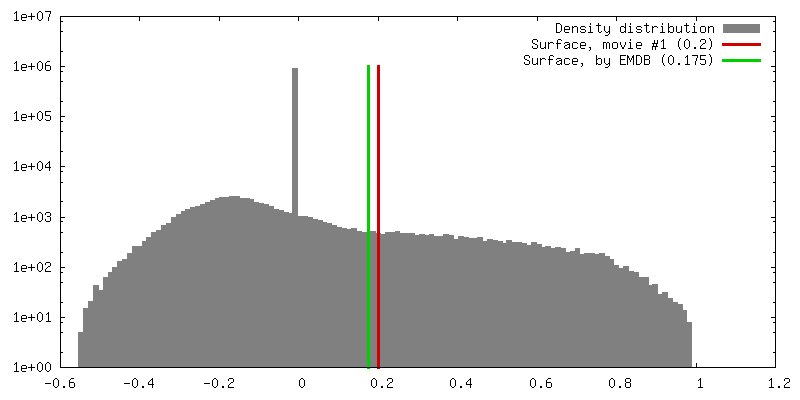
| Density |
| ||||||||||||||||||||||||||||||||||||||||||||||||||||||||||||||||||||
| Symmetry | Space group: 1 | ||||||||||||||||||||||||||||||||||||||||||||||||||||||||||||||||||||
| Details | EMDB XML:
CCP4 map header:
| ||||||||||||||||||||||||||||||||||||||||||||||||||||||||||||||||||||
 Z (Sec.)
 Y (Row.)
 X (Col.)


-Supplemental data
- Sample components
-Entire : MDA5-dsRNA
| Entire | Name: MDA5-dsRNA |
|---|---|
| Components |
|
-Supramolecule #1000: MDA5-dsRNA
| Supramolecule | Name: MDA5-dsRNA / type: sample / ID: 1000 / Number unique components: 2 |
|---|
-Macromolecule #1: MDA5
| Macromolecule | Name: MDA5 / type: protein_or_peptide / ID: 1 / Recombinant expression: Yes |
|---|---|
| Source (natural) | Organism: |
| Recombinant expression | Organism:  |
-Experimental details
-Structure determination
 Processing Processing | helical reconstruction |
|---|---|
| Aggregation state | filament |
-Sample preparation
| Vitrification | Cryogen name: NONE / Instrument: OTHER |
|---|
- Electron microscopy
| Microscope | FEI TECNAI 12 |
|---|---|
| Date | May 1, 2012 |
| Image recording | Category: FILM / Film or detector model: KODAK SO-163 FILM / Digitization - Scanner: NIKON COOLSCAN / Digitization - Sampling interval: 4.16 µm / Bits/pixel: 14 |
| Electron beam | Acceleration voltage: 80 kV / Electron source: TUNGSTEN HAIRPIN |
| Electron optics | Illumination mode: FLOOD BEAM / Imaging mode: BRIGHT FIELD |
| Sample stage | Specimen holder model: OTHER |
-Image processing
| Details | The particles were processed using IHRSR |
|---|---|
| Final reconstruction | Applied symmetry - Helical parameters - Δz: 43.6 Å Applied symmetry - Helical parameters - Δ&Phi: 82.7 ° Algorithm: OTHER / Resolution.type: BY AUTHOR / Resolution: 20.0 Å / Resolution method: FSC 0.5 CUT-OFF / Software - Name: Spider |
