Movie
 Controller


[English] 日本語
 Yorodumi
- EMDB-54301: Egalitarian-BicD in complex with ILS of I-factor mRNA (Structure B) -
+ Open data

- Basic information
| Entry |  | |||||||||
|---|---|---|---|---|---|---|---|---|---|---|
| Title | Egalitarian-BicD in complex with ILS of I-factor mRNA (Structure B) | |||||||||
 Map data | unsharpened map | |||||||||
 Sample |
| |||||||||
 Keywords | Complex / Transport / Localization / RNA BINDING PROTEIN | |||||||||
| Biological species |   Drosophila melanogaster (fruit fly) | |||||||||
| Method | single particle reconstruction / cryo EM / Resolution: 3.7 Å | |||||||||
 Authors | Singh K / Chilaeva S / McClintock MA / Bullock SL / Carter AP | |||||||||
| Funding support |  United Kingdom, European Union, 2 items
| |||||||||
 Citation | Journal: Nat Struct Mol Biol / Year: 2026 Title: Structural basis for recognition of diverse localizing mRNAs by Egl-BicD. Authors: Kashish Singh / Sabila Chilaeva / Mark A McClintock / Andrew P Carter / Simon L Bullock / Abstract: Localization of mRNAs is a widespread mechanism for dictating where proteins operate in cells and underpins many fundamental processes, from embryonic patterning to synaptic plasticity. This spatial ...Localization of mRNAs is a widespread mechanism for dictating where proteins operate in cells and underpins many fundamental processes, from embryonic patterning to synaptic plasticity. This spatial control is mediated by the interaction of 'localization signals' in target mRNAs with RNA-binding proteins (RBPs). These signals frequently lack overt sequence or structural patterns, raising the question of how specificity is achieved. Here we investigate this issue using the Drosophila RBP Egalitarian (Egl), which couples mRNAs to microtubule-based transport through Bicaudal D (BicD) and the dynein motor. We present cryo-electron microscopy structures of Egl-BicD bound to six different RNAs. Egl uses multiple noncanonical double-stranded RNA-binding domains to cooperatively form a recognition pocket around localization signals. Despite substantial variation in length and sequence, each signal adopts a bent stem-loop conformation that, together with base-pair identities at two defined sites, drives Egl engagement. We further demonstrate that Egl dimers couple RNA binding to transport initiation through coincident detection of two RNA elements within the same transcript. Thus, localizing mRNAs are recognized through a combination of shape, positional sequence features and number of structured RNA elements. This work reveals a molecular strategy by which diverse mRNAs can be selectively engaged by a single RBP. | |||||||||
| History |
|
- Structure visualization
| Supplemental images |
|---|

- Downloads & links
-EMDB archive
| Map data | emd_54301.map.gz | 89.2 MB |  EMDB map data format | |
|---|---|---|---|---|
| Header (meta data) | emd-54301-v30.xmlemd-54301.xml | 19.5 KB 19.5 KB | Display Display | EMDB header |
| FSC (resolution estimation) | emd_54301_fsc.xml | 12.8 KB | Display | FSC data file |
| Images |  emd_54301.png | 53.6 KB | ||
| Filedesc metadata | emd-54301.cif.gz | 4.6 KB | ||
| Others | emd_54301_additional_1.map.gzemd_54301_half_map_1.map.gzemd_54301_half_map_2.map.gz | 165 MB 165 MB 165 MB | ||
| Archive directory |  https://data.pdbj.org/pub/emdb/structures/EMD-54301ftp://data.pdbj.org/pub/emdb/structures/EMD-54301 | HTTPS FTP |
-Related structure data
| Related structure data |  9rvyC  9rvzC  9rw0C  9rw1C  9rw2C  9rw3C  9rw4C  9rw5C  9rw6C  9rw7C  9rw8C  9rw9C  9rwaC  9rwbC C: citing same article ( |
|---|


-Links
| EMDB pages | EMDB (EBI/PDBe) / EMDataResource |
|---|
-Map
| File | Download / File: emd_54301.map.gz / Format: CCP4 / Size: 178 MB / Type: IMAGE STORED AS FLOATING POINT NUMBER (4 BYTES) | ||||||||||||||||||||||||||||||||||||
|---|---|---|---|---|---|---|---|---|---|---|---|---|---|---|---|---|---|---|---|---|---|---|---|---|---|---|---|---|---|---|---|---|---|---|---|---|---|
| Annotation | unsharpened map | ||||||||||||||||||||||||||||||||||||
| Projections & slices | Image control
Images are generated by Spider. | ||||||||||||||||||||||||||||||||||||
| Voxel size | X=Y=Z: 1.059 Å | ||||||||||||||||||||||||||||||||||||
| Density |
| ||||||||||||||||||||||||||||||||||||
| Symmetry | Space group: 1 | ||||||||||||||||||||||||||||||||||||
| Details | EMDB XML:
|
 Z (Sec.)
 Y (Row.)
 X (Col.)


-Supplemental data
-Additional map: sharpened map
| File | emd_54301_additional_1.map | ||||||||||||
|---|---|---|---|---|---|---|---|---|---|---|---|---|---|
| Annotation | sharpened map | ||||||||||||
| Projections & Slices |
| ||||||||||||
| Density Histograms |


-Half map: #1
| File | emd_54301_half_map_1.map | ||||||||||||
|---|---|---|---|---|---|---|---|---|---|---|---|---|---|
| Projections & Slices |
| ||||||||||||
| Density Histograms |


-Half map: #2
| File | emd_54301_half_map_2.map | ||||||||||||
|---|---|---|---|---|---|---|---|---|---|---|---|---|---|
| Projections & Slices |
| ||||||||||||
| Density Histograms |


- Sample components
-Entire : Egalitarian-BicD in complex with ILS of I-factor mRNA
| Entire | Name: Egalitarian-BicD in complex with ILS of I-factor mRNA |
|---|---|
| Components |
|
-Supramolecule #1: Egalitarian-BicD in complex with ILS of I-factor mRNA
| Supramolecule | Name: Egalitarian-BicD in complex with ILS of I-factor mRNA / type: complex / ID: 1 / Parent: 0 / Macromolecule list: #1-#4 Details: Egalitarian-BicD in complex with I-factor localization signal (ILS) of I-factor retrotransposon mRNA |
|---|---|
| Source (natural) | Organism: Drosophila melanogaster (fruit fly) |
-Experimental details
-Structure determination
| Method | cryo EM |
|---|---|
 Processing | single particle reconstruction |
| Aggregation state | particle |
-Sample preparation
| Buffer | pH: 7.3 Details: 25 mM HEPES pH 7.3, 150 mM KCl, 1 mM MgCl2, 1mM DTT, 0.00125% IGEPAL |
|---|---|
| Vitrification | Cryogen name: ETHANE |
- Electron microscopy
| Microscope | TFS KRIOS |
|---|---|
| Image recording | Film or detector model: GATAN K3 BIOQUANTUM (6k x 4k) / Average electron dose: 48.0 e/Å2 |
| Electron beam | Acceleration voltage: 300 kV / Electron source:  FIELD EMISSION GUN |
| Electron optics | C2 aperture diameter: 100.0 µm / Illumination mode: FLOOD BEAM / Imaging mode: BRIGHT FIELD / Cs: 2.7 mm / Nominal defocus max: 3.5 µm / Nominal defocus min: 1.0 µm / Nominal magnification: 81000 |
| Experimental equipment | 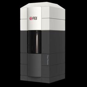 Model: Titan Krios / Image courtesy: FEI Company |