 Movie
Movie Controller

[English] 日本語
 Yorodumi
- EMDB-53938: Structure of the RhGB07 Prefusion, Closed-state trimeric spike protein -
+ Open data

- Basic information
| Entry |  | |||||||||
|---|---|---|---|---|---|---|---|---|---|---|
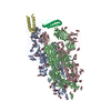
| Title | Structure of the RhGB07 Prefusion, Closed-state trimeric spike protein | |||||||||
 Map data Map data | ||||||||||
 Sample Sample |
| |||||||||
 Keywords Keywords | Spike protein of RhGB07 coronavirus / VIRAL PROTEIN | |||||||||
| Biological species |  Sarbecovirus sp. RhGB07 Sarbecovirus sp. RhGB07 | |||||||||
| Method | single particle reconstruction / cryo EM / Resolution: 2.97 Å | |||||||||
 Authors Authors | Lau K / Dong CN / Ekundayo B | |||||||||
| Funding support |  Switzerland, 1 items Switzerland, 1 items
| |||||||||
 Citation Citation | Journal: Biorxiv / Year: 2025 Title: Understanding the future risk of bat coronavirus spillover into humans - correlating sarbecovirus receptor usage, host range, and antigenicity Authors: Thakur N / Newman J / Ni D / Seow J / Hay AL / Li Y / Upadhyay A / Ekundayo BE / Peacock TP / Lau K / Doores KJ / Bailey D | |||||||||
| History |
|
- Structure visualization
| Supplemental images |
|---|

- Downloads & links
-EMDB archive
| Map data | emd_53938.map.gz | 52.3 MB |  EMDB map data format EMDB map data format | |
|---|---|---|---|---|
| Header (meta data) | emd-53938-v30.xmlemd-53938.xml | 18.6 KB 18.6 KB | Display Display | EMDB header |
| FSC (resolution estimation) | emd_53938_fsc.xml | 9.8 KB | Display | FSC data file |
| Images |  emd_53938.png emd_53938.png | 111 KB | ||
| Filedesc metadata | emd-53938.cif.gz | 6.9 KB | ||
| Others | emd_53938_half_map_1.map.gzemd_53938_half_map_2.map.gz | 95.6 MB 95.6 MB | ||
| Archive directory |  http://ftp.pdbj.org/pub/emdb/structures/EMD-53938ftp://ftp.pdbj.org/pub/emdb/structures/EMD-53938 http://ftp.pdbj.org/pub/emdb/structures/EMD-53938ftp://ftp.pdbj.org/pub/emdb/structures/EMD-53938 | HTTPS FTP |
-Related structure data
-Links
| EMDB pages | EMDB (EBI/PDBe) / EMDataResource |
|---|
-Map
| File | Download / File: emd_53938.map.gz / Format: CCP4 / Size: 103 MB / Type: IMAGE STORED AS FLOATING POINT NUMBER (4 BYTES) | ||||||||||||||||||||||||||||||||||||
|---|---|---|---|---|---|---|---|---|---|---|---|---|---|---|---|---|---|---|---|---|---|---|---|---|---|---|---|---|---|---|---|---|---|---|---|---|---|
| Projections & slices | Image control
Images are generated by Spider. | ||||||||||||||||||||||||||||||||||||
| Voxel size | X=Y=Z: 1.38333 Å | ||||||||||||||||||||||||||||||||||||
| Density |
| ||||||||||||||||||||||||||||||||||||
| Symmetry | Space group: 1 | ||||||||||||||||||||||||||||||||||||
| Details | EMDB XML:
|
 Z (Sec.)
 Y (Row.)
 X (Col.)


-Supplemental data
-Half map: #2
| File | emd_53938_half_map_1.map | ||||||||||||
|---|---|---|---|---|---|---|---|---|---|---|---|---|---|
| Projections & Slices |
| ||||||||||||
| Density Histograms |


-Half map: #1
| File | emd_53938_half_map_2.map | ||||||||||||
|---|---|---|---|---|---|---|---|---|---|---|---|---|---|
| Projections & Slices |
| ||||||||||||
| Density Histograms |


- Sample components
-Entire : Trimeric RhGB07 Spike
| Entire | Name: Trimeric RhGB07 Spike |
|---|---|
| Components |
|
-Supramolecule #1: Trimeric RhGB07 Spike
| Supramolecule | Name: Trimeric RhGB07 Spike / type: complex / ID: 1 / Parent: 0 / Macromolecule list: #1 |
|---|---|
| Source (natural) | Organism: Sarbecovirus sp. RhGB07 |
-Macromolecule #1: RhGB07 Spike- T4 foldon fusion protein
| Macromolecule | Name: RhGB07 Spike- T4 foldon fusion protein / type: protein_or_peptide / ID: 1 / Number of copies: 3 / Enantiomer: LEVO |
|---|---|
| Source (natural) | Organism: Sarbecovirus sp. RhGB07 |
| Molecular weight | Theoretical: 138.592109 KDa |
| Recombinant expression | Organism:   Cricetulus griseus (Chinese hamster) Cricetulus griseus (Chinese hamster) |
| Sequence | String: DGCGRLSQKT QPKLEQFNSS RRGLYYFDDI FRSSLRVLTS GYFLPFGSNV TRYEVKQVPQ VPIYYDNPLV PFGHGVYFAV TEKSNVVRG WIFGSTMDNT TQSAVLVNNG THIVVNVCNF NFCADPMFAV SAGQSYKSWI YTSSTNCTYF QAFPFAIETR P NGTGPFTH ...String: DGCGRLSQKT QPKLEQFNSS RRGLYYFDDI FRSSLRVLTS GYFLPFGSNV TRYEVKQVPQ VPIYYDNPLV PFGHGVYFAV TEKSNVVRG WIFGSTMDNT TQSAVLVNNG THIVVNVCNF NFCADPMFAV SAGQSYKSWI YTSSTNCTYF QAFPFAIETR P NGTGPFTH LREHVFRNVD GFLHVYHNYE VINVQNGLPS GFSVLRPIFK LPLGLNITYI KAVMTLFSST TSNFIAAVAS LH YVGYLQP RTMLFHFDQN GTIVDAIDCS QNPLSELKCT TKSFSLEKGI YQTSNFRVTP TTEVIRFPNV TQLCPFSDVF NRT KFPSVY AWERMSIRDC VADYAVLYNS SISFSTFECY GVSPTKLNDL CFSSVYADYF VVRGDDVKQI APGKTGVIAD YNYK LPDDF VGCVLAWNSR HVDSKQGFYY RLFRHGKIKP YQRDMSRELY SPSGTPCTTE GFNCYDPLAH YAFASPGIGT GYQPF RVVV LSFQLLNAPA TVCGPKQSTD LVKNKCVNFN FNGLTGTGVL TDSVKKFQPF QQFGRDSSDF TDSVKDPKTL EILDIT PCS YGGVSVITPG TNTSDAVAVL YQDVNCTDVP TMLHIDQVSS DWRVYAANTG GNMFQTQAGC LVGATYDNTS YECDIPV GA GVCAKFQINT GASQSSILAY TMSLGEDSNV AYSNNSIAIP TNFSISVTTE VLPVSMTKTS VDCNMYICGD STECSSLL L QYGSFCQQLN RALAGVSVEQ DKNTQDVFAQ VKSIYKVPAI KDFGGFNFSH ILPDPAKPSK RSFIEDLLYN KVTLADPGF MKQYGDCLGG VNARDLICAQ KFNGLTVLPP LLTDDMIAAY TSALVSGTAL SGYTFGAGAA LQIPFAMQMA YRFNGIGVTQ NVLYENQKQ IANQFNKAIS QIQESLTTTA TALGKLQDVI NQNAIALNTL VKQLSSNFGA ISSVLNDILS RLDPPEAEVQ I DRLITGRL QSLQTYVTQQ LIRAAEIRAS ANLAATKMSE CVLGQSKRVD FCGKGYHLMS FPQAAPHGVV FLHVTYVPSQ EQ NFTTAPA ICHKGKAHFP REGVFVTNGT HWFVTQRNFY QPEVITTENT FESGNCDVVI GIVNNTVYDP LQPELESFKE ELD KYFKNH TSPEVGFDDI SGINASVVDI KKEIAHLNEI ASKLNESLID LQELGKYEQG SGYIPEAPRD GQAYVRKDGE WVLL STFLG RSLEVLFQGP GHHHHHHHHS AWSHPQFEKG GGSGGGGSGG SAWSHPQFEK |
-Macromolecule #6: 2-acetamido-2-deoxy-beta-D-glucopyranose
| Macromolecule | Name: 2-acetamido-2-deoxy-beta-D-glucopyranose / type: ligand / ID: 6 / Number of copies: 18 / Formula: NAG |
|---|---|
| Molecular weight | Theoretical: 221.208 Da |
| Chemical component information |  ChemComp-NAG: |
-Experimental details
-Structure determination
| Method | cryo EM |
|---|---|
 Processing Processing | single particle reconstruction |
| Aggregation state | particle |
-Sample preparation
| Concentration | 2 mg/mL |
|---|---|
| Buffer | pH: 7.5 |
| Grid | Model: Quantifoil R1.2/1.3 / Material: GOLD / Mesh: 400 / Support film - Material: CARBON / Support film - topology: HOLEY / Pretreatment - Type: GLOW DISCHARGE / Pretreatment - Time: 120 sec. |
| Vitrification | Cryogen name: ETHANE |
- Electron microscopy
| Microscope | TFS KRIOS |
|---|---|
| Software | Name: EPU |
| Image recording | Film or detector model: FEI FALCON IV (4k x 4k) / Average exposure time: 5.04 sec. / Average electron dose: 60.0 e/Å2 |
| Electron beam | Acceleration voltage: 300 kV / Electron source:  FIELD EMISSION GUN FIELD EMISSION GUN |
| Electron optics | C2 aperture diameter: 50.0 µm / Illumination mode: FLOOD BEAM / Imaging mode: BRIGHT FIELD / Nominal defocus max: 2.4 µm / Nominal defocus min: 1.0 µm / Nominal magnification: 96000 |
| Sample stage | Specimen holder model: FEI TITAN KRIOS AUTOGRID HOLDER / Cooling holder cryogen: NITROGEN |
| Experimental equipment |  Model: Titan Krios / Image courtesy: FEI Company |
+Image processing

-Atomic model buiding 1
| Initial model | Chain - Source name: AlphaFold / Chain - Initial model type: in silico model |
|---|---|
| Software | Name: PHENIX / Details: EMplace |
| Details | Model was docked using Phenix EMPlace followed by manual building in Coot and phenix real space refine |
| Refinement | Space: REAL |
| Output model |  PDB-9rdt: |
